

![Fibronectin antibody [N1N2], N-term产品图](https://img1.dxycdn.com/2023/0711/255/2344116916935858761-14.jpg)
- ¥1700 - 4000
- GeneTex

- 美国
- GTX112794
- 2025年07月10日
- WB, ICC/IF, IHC-P, IHC-Fr, IP, ELISA
- Rabbit
- Human, Mouse, Rat

企业认证
相关产品推荐更多 >
![MUC1 antibody [C595 (NCRC48)] (FITC)](https://img1.dxycdn.com/2023/0711/451/6106825198874858761-14.jpg!wh200)


万千商家帮你免费找货
0 人在求购买到急需产品
- 详细信息
- 文献和实验
- 技术资料
- 免疫原:
Recombinant protein encompassing a sequence within the N-terminus region of human Fibronectin. The exact sequence is proprietary.
- 亚型:
IgG
- 形态:
Liquid
- 保存条件:
Store as concentrated solution. Centrifuge briefly prior to opening vial. For short-term storage (1-2 weeks), store at 4ºC. For long-term storage, aliquot and store at -20ºC or below. Avoid multiple freeze-thaw cycles.
- 克隆性:
Polyclonal
- 标记物:
Unconjugated
- 适应物种:
Human, Mouse, Rat
- 保质期:
12 months from the shipping date of the product.
- 抗原来源:
Human
- 目录编号:
GTX112794
- 级别:
Primary Antibodies
- 库存:
Available
- 供应商:
GeneTex
- 宿主:
Rabbit
- 应用范围:
WB, ICC/IF, IHC-P, IHC-Fr, IP, ELISA
- 浓度:
0.34 mg/ml (Please refer to the vial label for the specific concentration.)
- 靶点:
Fibronectin
- 抗体英文名:
Fibronectin antibody [N1N2], N-term
- 抗体名:
Fibronectin 抗体 [N1N2], N-term
- 规格:
100 μl/25 μl
| 规格: | 100 μl | 产品价格: | ¥4000.0 |
|---|---|---|---|
| 规格: | 25 μl | 产品价格: | ¥1700.0 |
Fibronectin antibody [N1N2], N-term detects Fibronectin protein at cell membrane by immunohistochemical analysis.
Sample: Paraffin-embedded mouse esophagus.
Fibronectin stained by Fibronectin antibody [N1N2], N-term (GTX112794) diluted at 1:500.
Antigen Retrieval: Citrate buffer, pH 6.0, 15 min
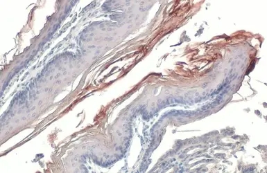
Fibronectin antibody [N1N2], N-term detects Fibronectin protein at cell membrane by immunohistochemical analysis.
Sample: Paraffin-embedded rat colon.
Fibronectin stained by Fibronectin antibody [N1N2], N-term (GTX112794) diluted at 1:500.
Antigen Retrieval: Citrate buffer, pH 6.0, 15 min
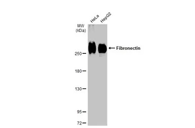
Various whole cell extracts (30 μg) were separated by 5% SDS-PAGE, and the membrane was blotted with Fibronectin antibody [N1N2], N-term (GTX112794) diluted at 1:1000. The HRP-conjugated anti-rabbit IgG antibody (GTX213110-01) was used to detect the primary antibody.
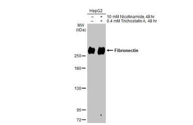
Untreated (–) and treated (+) HepG2 whole cell extracts (30 μg) were separated by 5% SDS-PAGE, and the membrane was blotted with Fibronectin antibody [N1N2], N-term (GTX112794) diluted at 1:1000. The HRP-conjugated anti-rabbit IgG antibody (GTX213110-01) was used to detect the primary antibody.
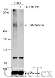
Non-transfected (–) and transfected (+) HeLa whole cell extracts (30 μg) were separated by 5% SDS-PAGE, and the membrane was blotted with Fibronectin antibody [N1N2], N-term (GTX112794) diluted at 1:2000. The HRP-conjugated anti-rabbit IgG antibody (GTX213110-01) was used to detect the primary antibody, and the signal was developed with Trident ECL plus-Enhanced.
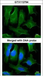
Immunofluorescence analysis of PFA-fixed HeLa, using Fibronectin (GTX112794) antibody at 1:200 dilution.
Fibronectin antibody [N1N2] detects Fibronectin protein by western blot analysis. Mouse tissue extracts (50 μg) was separated by 5% SDS-PAGE, and the membrane was blotted with Fibronectin antibody [N1N2] (GTX112794) diluted at 1:1000. The HRP-conjugated anti-rabbit IgG antibody (GTX213110-01) was used to detect the primary antibody.
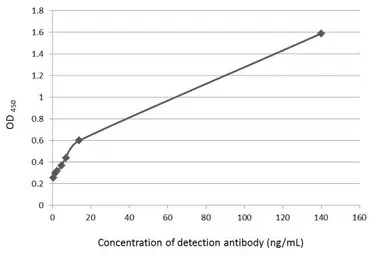
Indirect ELISA analysis was performed by coating plate with 100 μL of recombinant Fibronectin protein at concentration of 10 μg/mL. The coated protein is detected with Fibronectin antibody [N1N2], N-term (GTX112794) at rangeing from 0.5 to 140 ng/mL.
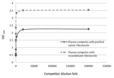
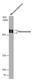
Competitive ELISA detection of human fibronectin in plasma using GTX112794. Recombinant or purified native Fibronectin protein was coated to an ELISA plate at concentration of 10 μg/ml. Human plasma was applied as the competitors and was serial diluted at 1, 20, 200, 2000, 20000, 200000X dilutions. The primary antibody concentration was diluted to 14 ng/mL. Rabbit IgG antibody (HRP) (GTX213110-01) was diluted at 1:2,000 and used to detect the primary antibody.
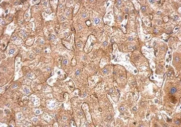
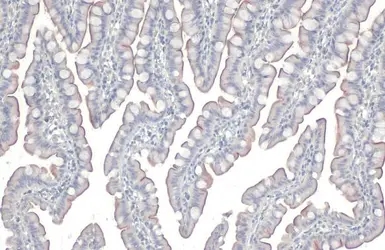
Fibronectin antibody [N1N2], N-term detects FN1 protein at cytosol on human hepatoma by immunohistochemical analysis.
Sample: Paraffin-embedded hepatoma.
Fibronectin antibody [N1N2], N-term (GTX112794) dilution: 1:500.
Antigen Retrieval: Trilogy™ (EDTA based, pH 8.0) buffer, 15min
风险提示:丁香通仅作为第三方平台,为商家信息发布提供平台空间。用户咨询产品时请注意保护个人信息及财产安全,合理判断,谨慎选购商品,商家和用户对交易行为负责。对于医疗器械类产品,请先查证核实企业经营资质和医疗器械产品注册证情况。
 文献和实验
文献和实验Chen LC et al., Cells 2021 (PMID:34685727)
Gaur G et al., Antibiotics (Basel) 2021 (PMID:34827233)
Wang LT et al., Cancer Lett 2021 (PMID:34265398)
Hsu TW et al., Biomaterials 2021 (PMID:33780686)
Altera A et al., Graefes Arch Clin Exp Ophthalmol 2021 (PMID:33760980)
Chang CJ et al., Biochem Pharmacol 2021 (PMID:33285108)
Chang YX et al., Cancers (Basel) 2020 (PMID:33371405)
Horn LA et al., J Immunother Cancer 2020 (PMID:32188703)
Wang LT et al., EMBO Rep 2020 (PMID:31908141)
Yokdang N et al., Oncogene 2016 (PMID:26387542)
Mohankumar K et al., Endocrinology 2020 (PMID:32099996)
Zhang L et al., Cells 2019 (PMID:31640200)
Hung KF et al., Int J Biol Sci 2019 (PMID:31182927)
Wang HC et al., Int J Mol Sci 2019 (PMID:31438571)
Li-Chien Chang et al., Nutrition 2019 (Epub)
Kuo YL et al., Sci Rep 2019 (PMID:31311941)
Kai-Feng Hung et al., Int. J. Biol. Sci. 2019;15(5)
Miki F et al., Biol Reprod 2019 (PMID:30649202)
Chu CA et al., EBioMedicine 2019 (PMID:30982765)
Pei YF et al., Am J Pathol 2019 (PMID:30735628)
Tsai CH et al., Materials (Basel) 2019 (PMID:30634440)
Hou YY et al., Oncol Lett 2016 (PMID:27313732)
Chung IH et al., Oncotarget 2016 (PMID:26840566)
Luo CW et al., Exp Cell Res 2018 (PMID:29305962)
Pon JR et al., Nat Commun 2015 (PMID:26245647)
Dominguez C et al., JCI Insight 2017 (PMID:29093275)
Lin EY et al., Environ Toxicol 2017 (PMID:29064158)
Pei YF et al., Biochim Biophys Acta 2017 (PMID:29045811)
Chou HC et al., Toxicological Sciences 2017 (Epub)
Zhang X et al., Cell Biol Int 2017 (PMID:28699302)
Chao CC et al., FEBS Lett 2017 (PMID:28542779)
Zhao B et al., J Proteomics 2015 (PMID:25173099)
Schendel SA et al., Aesthet Surg J 2017 (PMID:28510634)
Wang LT et al., Oncogene 2017 (PMID:28368406)
Sorrentino G et al., Nat Commun 2017 (PMID:28102225)
Chen S et al., World J Urol 2016 (PMID:28013345)
Watanabe R et al., JACC Basic Transl Sci 2016;1(6)
Dominguez C et al., Cell Death Dis 2016 (PMID:27685624)
Cheng YL et al., J. Mater. Chem. B. 2016 (38)
Sridaran D et al., Tumour Biol 2016 (PMID:27460079)
Yamamoto K et al., Peptides 2016 (PMID:27346255)
Hamilton DH et al., Clin Cancer Res 2016 (PMID:27267852)
Xu M et al., Oncotarget 2015 (PMID:25460509)
Wang HL et al., J Cell Sci 2015 (PMID:26359301)
Julia Pon et al., 0 2010
N Yokdang et al., Oncogene 2015
Atik E et al., Indian J Cancer 2014 (PMID:24947099)
Yang W et al., J Biomed Mater Res A 2013 (PMID:24178590)
Insalaco L et al., J Cell Mol Med 2012 (PMID:22260151)
Li YJ et al., iScience 2023 (PMID:37138780)
Chua HH et al., Cell Mol Gastroenterol Hepatol 2023 (PMID:36191855)
Chen H et al., Regen Biomater 2023 (PMID:36683749)
Su MT et al., Pharmaceuticals (Basel) 2022 (PMID:36558974)
Chiou WC et al., Biomed Pharmacother 2023 (PMID:36906971)
Catarinella G et al., Cell Death Dis 2022 (PMID:36028501)
Zhao W et al., Bioeng Transl Med 2022 (PMID:36176617)
Lee WJ et al., J Food Drug Anal 2021 (PMID:35696218)
Chi-Wen Luo et al., Am J Cancer Res 2020 (PMID:33415002)
In the field of therapeutic recombinant proteins, monoclonal antibodies (mAbs) have achieved a rising success with more than 30 mAbs that have reached the market in the past 20 years. From a structural standpoint, one of the most important
to protect the antibody. These should be stored in smaller aliquots (about 1 ml) and frozen for long term storage though the protected solution is also stable for months at four degrees. Ascites fluid: This is a bit more problematic as it may contain
Materials 0.1M NaHC03 pH9 DMSO NHS-Biotin (N-Hydroxysuccinimidobiotin, Sigma #H-1759) PBS Procedure Dialyze the sample against carbonate buffer. After dialysis, adjust
 技术资料
技术资料暂无技术资料 索取技术资料

