


- ¥1700 - 4000
- GeneTex

- 美国
- GTX124252
- 2025年07月12日
- WB, ICC/IF, IHC-P, FACS, IP, IHC
- Rabbit
- Dengue virus

企业认证
相关产品推荐更多 >


![STEAP1 antibody [J2D2]](https://img1.dxycdn.com/2023/0711/451/6106825198874858761-14.jpg!wh200)
![CD22 antibody [OX-97] (Biotin)](https://custom.dxycdn.com/trademd/upload/pic/2011/12/14/1323695851.jpg!small.t.1)

万千商家帮你免费找货
0 人在求购买到急需产品
- 详细信息
- 文献和实验
- 技术资料
- 免疫原:
Recombinant protein encompassing a sequence within the center region of Dengue virus NS3 protein (Dengue virus 2 (strain 16681 PDK 53)). The exact sequence is proprietary.
- 亚型:
IgG
- 形态:
Liquid
- 保存条件:
Store as concentrated solution. Centrifuge briefly prior to opening vial. For short-term storage (1-2 weeks), store at 4ºC. For long-term storage, aliquot and store at -20ºC or below. Avoid multiple freeze-thaw cycles.
- 克隆性:
Polyclonal
- 标记物:
Unconjugated
- 适应物种:
Dengue virus
- 保质期:
12 months from the shipping date of the product.
- 抗原来源:
Dengue virus
- 目录编号:
GTX124252
- 级别:
Primary Antibodies
- 库存:
Available
- 供应商:
GeneTex
- 宿主:
Rabbit
- 应用范围:
WB, ICC/IF, IHC-P, FACS, IP, IHC
- 浓度:
0.15 mg/ml (Please refer to the vial label for the specific concentration.)
- 靶点:
Dengue virus NS3 protein
- 抗体英文名:
Dengue virus NS3 protein antibody
- 抗体名:
Dengue virus NS3 protein 抗体
- 规格:
100 μl/25 μl
| 规格: | 100 μl | 产品价格: | ¥4000.0 |
|---|---|---|---|
| 规格: | 25 μl | 产品价格: | ¥1700.0 |
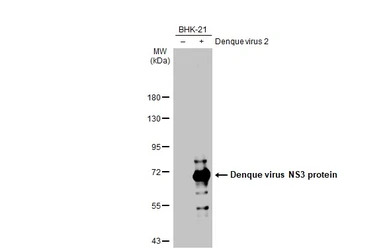
Non-infected (–) and infected (+) BHK-21 whole cell extracts (15 μg) were separated by 7.5% SDS-PAGE, and the membrane was blotted with Dengue virus NS3 protein antibody (GTX124252) diluted at 1:2000. The HRP-conjugated anti-rabbit IgG antibody (GTX213110-01) was used to detect the primary antibody.
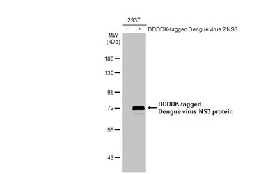
Non-transfected (–) and transfected (+) 293T whole cell extracts (30 μg) were separated by 7.5% SDS-PAGE, and the membrane was blotted with Dengue virus NS3 protein antibody (GTX124252) diluted at 1:5000. The HRP-conjugated anti-rabbit IgG antibody (GTX213110-01) was used to detect the primary antibody.
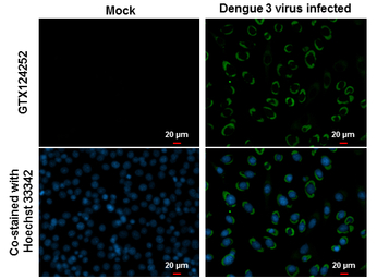
NS3 (Dengue virus) antibody detects NS3 (Dengue virus) protein at cytoplasm by immunofluorescent analysis.
Samples: BHK-21 cells mock (left) and infected with Dengue virus 3 (right) were fixed in MeOH.
Green: NS3 (Dengue virus) protein stained by NS3 (Dengue virus) antibody (GTX124252) diluted at 1:2000.
Blue: Hoechst 33342 staining.
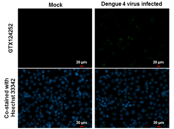
NS3 (Dengue virus) antibody detects NS3 (Dengue virus) protein at cytoplasm by immunofluorescent analysis.
Samples: BHK-21 cells mock (left) and infected with Dengue virus 4 (right) were fixed in MeOH.
Green: NS3 (Dengue virus) protein stained by NS3 (Dengue virus) antibody (GTX124252) diluted at 1:2000.
Blue: Hoechst 33342 staining.
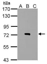
Sample (20 μg of whole cell lysate)
A: BHK-21
B: Dengue virus 2 infect BHK-21
C: JEV infect BHK-21
7.5% SDS PAGE
GTX124252 diluted at 1:2000
The HRP-conjugated anti-rabbit IgG antibody (GTX213110-01) was used to detect the primary antibody.
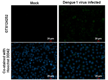
NS3 (Dengue virus) antibody detects NS3 (Dengue virus) protein at cytoplasm by immunofluorescent analysis.
Samples: BHK-21 cells mock (left) and infected with Dengue virus 1 (right) were fixed in MeOH.
Green: NS3 (Dengue virus) protein stained by NS3 (Dengue virus) antibody (GTX124252) diluted at 1:2000.
Blue: Hoechst 33342 staining.
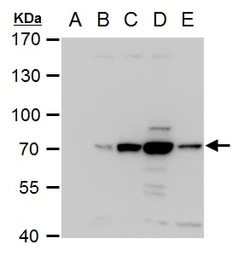
NS3 (Dengue virus) antibody detects NS3 (Dengue virus) protein by western blot analysis.
A. 30 μg BHK-21 whole cell extract
B. 30 μg whole cell extract of Dengue virus type 1 infected BHK-21 cells
C. 30 μg whole cell extract of Dengue virus type 2 infected BHK-21 cells
D. 30 μg whole cell extract of Dengue virus type 3 infected BHK-21 cells
E. 30 μg whole cell extract of Dengue virus type 4 infected BHK-21 cells
7.5% SDS-PAGE
NS3 (Dengue virus) antibody (GTX124252) dilution: 1:2000
The HRP-conjugated anti-rabbit IgG antibody (GTX213110-01) was used to detect the primary antibody.
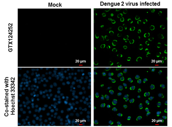
NS3 (Dengue virus) antibody detects NS3 (Dengue virus) protein at cytoplasm by immunofluorescent analysis.
Samples: BHK-21 cells mock (left) and infected with Dengue virus 2 (right) were fixed in MeOH.
Green: NS3 (Dengue virus) protein stained by NS3 (Dengue virus) antibody (GTX124252) diluted at 1:2000.
Blue: Hoechst 33342 staining.
风险提示:丁香通仅作为第三方平台,为商家信息发布提供平台空间。用户咨询产品时请注意保护个人信息及财产安全,合理判断,谨慎选购商品,商家和用户对交易行为负责。对于医疗器械类产品,请先查证核实企业经营资质和医疗器械产品注册证情况。
 文献和实验
文献和实验Bautista-Carbajal P et al., Virology 2017 (PMID:27940224)
Barratt-Boyes SM et al., Comp Immunol Microbiol Infect Dis 2012 (PMID:22293473)
Sun P et al., J Immunol Methods 2016 (PMID:27856192)
Wu YW et al., Sci Rep 2016 (PMID:27558165)
Cheng YL et al., Sci Rep 2016 (PMID:27561946)
Tsai TT et al., Sci Rep 2016 (PMID:27279150)
Morales L et al., Biomedica 2016 37()
Marceau C.D. et al., Nature 2016 (PMID:27383987)
Yu CY et al., PLoS Pathog 2015 (PMID:26717518)
Chu YT et al., Immunology 2015 (PMID:26059780)
Naik NG et al., J Virol 2015 (PMID:25878113)
Wan SW et al., PLoS One 2014 (PMID:24658118)
Angel-Ambrocio AH et al., Virus Res 2015 (PMID:25598317)
Sarathy VV et al., J Virol 2014 (PMID:25392217)
Soto-Acosta R et al., Antiviral Res 2014 (PMID:25017471)
Wu CW et al., Patent. 2012
Pan P et al., Signal Transduct Target Ther 2023 (PMID:37160897)
Han Y et al., Cell Stem Cell 2022 (PMID:36206731)
Hu M et al., Viruses 2022 (PMID:36146879)
Wei KC et al., Microbiol Spectr 2023 (PMID:36629424)
Udawatte DJ et al., Front Cell Infect Microbiol 2022 (PMID:36310878)
Yousefi M et al., PLoS Pathog 2022 (PMID:35939522)
Shwetha Shivaprasad et al., PLoS Pathog 2022 (PMID:35482886)
Thippayawan Ratanakomol et al., BMC Res Notes 2022 (PMID:35509105)
Vinit Upasani et al., Front Immunol 2020 (PMID:33643283)
Yen-Chen Chen et al., Pharmaceuticals (Basel) 2021 (PMID:33917182)
Carlos Noe Farfan-Morales et al., Sci Rep 2021 (PMID:33888740)
Pan Pan et al., PLoS Pathog 2021 (PMID:34310658)
Kongmanas K et al., PLoS Negl Trop Dis 2020 (PMID:33216752)
Brown RJP et al., Sci Adv 2020 (PMID:33148654)
Arias-Arias JL et al., J Biol Chem 2020 (PMID:31919100)
Zhou L et al., Antiviral Res 2021 (PMID:34801589)
Jhan MK et al., Cells 2021 (PMID:34831405)
Campos RK et al., Nucleic Acids Res 2020 (PMID:32890404)
Rathore APS et al., Front Immunol 2021 (PMID:34168651)
Aviner R et al., Nature 2021 (PMID:34408324)
Burke JM et al., Sci Adv 2021 (PMID:34088676)
Aguilar-Brise?o JA et al., Nat Commun 2020 (PMID:32576819)
Palacios-R?palo SN et al., Arch Virol 2021 (PMID:33682072)
Lin JJ et al., PLoS Pathog 2021 (PMID:33784371)
Tammineni ER et al., Virology 2021 (PMID:33249258)
Liu B et al., Virol Sin 2020 (PMID:33044659)
Vial T et al., Proc Natl Acad Sci U S A 2020 (PMID:33087565)
Pahmeier F et al., J Virol 2020 (PMID:33257477)
Pan P et al., Front Microbiol 2019 (PMID:31824450)
Lee YR et al., Kaohsiung J Med Sci 2020 (PMID:32783363)
Barbier V et al., Virology 2017 (PMID:27816895)
Afroz S et al., Front Immunol 2020 (PMID:32903536)
De Jes?s-Gonz?lez LA et al., Viruses 2020 (PMID:32466480)
Wei KC et al., Viruses 2020 (PMID:32121148)
Mohamed B et al., Sci Rep 2020 (PMID:32132633)
Butler, M et al., Viruses 2020 (PMID:32560467)
Wei KC et al., Front Cell Infect Microbiol 2018 (PMID:30186771)
Hitakarun A et al., Sci Rep 2020 (PMID:32001767)
Evans AS et al., J Virol 2020 (PMID:32102874)
Shrivastava G et al., Front Immunol 2020 (PMID:32210961)
P?rez-Olais JH et al., Virus Res 2019 (PMID:31626875)
Xie X et al., Cell Host Microbe 2019 (PMID:31631053)
Chuang FK et al., Antimicrob Agents Chemother 2019 (PMID:31636070)
Xing H et al., Virus Res 2020 (PMID:31676367)
Rothan HA et al., Antiviral Res 2019 (PMID:31421166)
Rafael K. Campos et al., bioRxiv 2019 (Epub)
Hammack C et al., J Virol 2019 (PMID:31375586)
Ooi YS et al., Nat Microbiol 2019 (PMID:31384002)
Nandan S. Gokhale et al., bioRxiv 2019 (Epub)
Frakolaki E et al., Cells 2018 (PMID:30513781)
Abernathy E et al., PLoS Biol 2019 (PMID:30608919)
Arias-Arias JL et al., Am J Trop Med Hyg 2018 (PMID:30398136)
Hishiki T et al., Front Microbiol 2017 (PMID:28912773)
Ramirez L et al., Virus Res 2018 (PMID:30278191)
Kao JC et al., PLoS Negl Trop Dis 2018 (PMID:30125275)
Juan Fidel Osuna-Ramos et al., bioRxiv 2018 (Epub)
Margot Cervantes-Salazar et al., bioRxiv 2018 (Epub)
Azia S Evans et al., bioRxiv 2018;2018(Epub)
Zhang J et al., Cell Host Microbe 2018 (PMID:29902443)
Lai YC et al., Antiviral Res 2018 (PMID:29752950)
Sarathy VV et al., Sci Rep 2018 (PMID:29559699)
McFadden MJ et al., Viruses 2018 (PMID:29495257)
Reyes-Ruiz JM et al., Virology 2017 (PMID:29272748)
Reid DW et al., J Virol 2018 (PMID:29321322)
Wu X et al., Cell 2017 (PMID:29249360)
St John AL et al., Elife 2013 (PMID:23638300)
Wan SW et al., J Immunol 2017 (PMID:28904127)
Lai YC et al., Sci Rep 2017 (PMID:28765561)
Morales L et al., Biomedica 2017 37()
Soto-Acosta R et al., PLoS Pathog 2017 (PMID:28384260)
Medin CL et al., J Virol Methods 2015 (PMID:25445884)
Zou J et al., J Virol 2015 (PMID:25589636)
Zou J et al., J Virol 2015 (PMID:25568208)
Campos RK et al., J Virol 2016 (PMID:27974556)
against dengue, timely diagnosis is therefore necessary for patient management. Laboratory diagnosis is carried out by virus isolation, demonstration of viral antigen, presence of viral nucleic acid, and antibodies. Further, recombinant dengue envelope protein
Fluorimetric and HPLC-Based Dengue Virus Protease Assays Using a FRET Substrate
The number of dengue virus infections is increasing and the dengue NS2B–NS3 protease is considered a promising target for the development of antiviral therapies. Therefore, reliable and fast screening systems are needed for the discovery
Characterization of the Dengue Virus Envelope Glycoprotein Expressed in Pichia pastoris
The full-length and truncated forms of recombinant envelope (E) glycoprotein from Dengue virus type 1, Singapore strain S275/90 were expressed in the yeast, Pichia pastoris , using a secretory vector. A truncated form of the E protein
 技术资料
技术资料暂无技术资料 索取技术资料

