


- ¥4000
- GeneTex

- 美国
- GTX124207
- 2026年02月26日
- WB, ICC/IF, IHC-P, IHC-Wm, IP, ChIP assay, IHC
- Rabbit
- Human, Mouse, Rat, Monkey

企业认证
相关产品推荐更多 >


![JAK2 antibody [1C1]](https://img1.dxycdn.com/2023/0711/451/6106825198874858761-14.jpg!wh200)
万千商家帮你免费找货
0 人在求购买到急需产品
- 详细信息
- 文献和实验
- 技术资料
- 免疫原:
Recombinant protein encompassing a sequence within the center region of human TET1. The exact sequence is proprietary.
- 亚型:
IgG
- 形态:
Liquid
- 保存条件:
Store as concentrated solution. Centrifuge briefly prior to opening vial. For short-term storage (1-2 weeks), store at 4ºC. For long-term storage, aliquot and store at -20ºC or below. Avoid multiple freeze-thaw cycles.
- 克隆性:
Polyclonal
- 标记物:
Unconjugated
- 适应物种:
Human, Mouse, Rat, Monkey
- 保质期:
12 months from the shipping date of the product.
- 抗原来源:
Human
- 目录编号:
GTX124207
- 级别:
Primary Antibodies
- 库存:
Available
- 供应商:
GeneTex
- 宿主:
Rabbit
- 应用范围:
WB, ICC/IF, IHC-P, IHC-Wm, IP, ChIP assay, IHC
- 浓度:
1.05 mg/ml (Please refer to the vial label for the specific concentration.)
- 靶点:
TET1
- 抗体英文名:
TET1 antibody [N3C1]
- 抗体名:
TET1 抗体 [N3C1]
- 规格:
100 μl
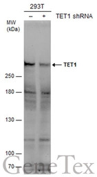
Non-transfected (–) and transfected (+) 293T whole cell extracts (50 μg) were separated by 5% SDS-PAGE, and the membrane was blotted with TET1 antibody [N3C1] (GTX124207) diluted at 1:1000. The HRP-conjugated anti-rabbit IgG antibody (GTX213110-01) was used to detect the primary antibody.
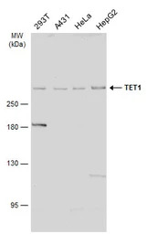
Various whole cell extracts (30 μg) were separated by 5% SDS-PAGE, and the membrane was blotted with TET1 antibody [N3C1] (GTX124207) diluted at 1:2000. The HRP-conjugated anti-rabbit IgG antibody (GTX213110-01) was used to detect the primary antibody.
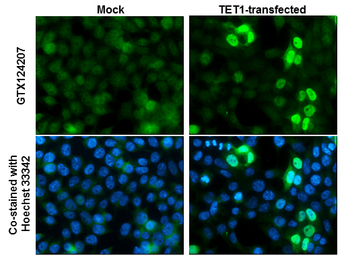
TET1 antibody [N3C1] detects TET1 protein at nucleus by immunofluorescent analysis.
Sample: Mock and transfected 293T cells were fixed in 4% PFA at RT for 15 min.
Green: TET1 stained by TET1 antibody [N3C1] (GTX124207) diluted at 1:1000.
Blue: Hoechst 33342 staining.
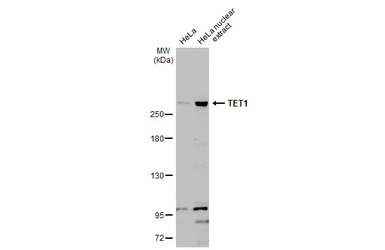
HeLa whole cell and nuclear extracts (30 μg) were separated by 5% SDS-PAGE, and the membrane was blotted with TET1 antibody [N3C1] (GTX124207) diluted at 1:1000. The HRP-conjugated anti-rabbit IgG antibody (GTX213110-01) was used to detect the primary antibody.
Immunohistochemical analysis of paraffin-embedded Hela xenograft, using TET1 (GTX124207) antibody at 1:1000 dilution.
Antigen Retrieval: Trilogy™ (EDTA based, pH 8.0) buffer, 15min
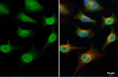
TET1 antibody [N3C1] detects TET1 protein at cytoplasm and nucleus by immunofluorescent analysis.
Sample: HeLa cells were fixed in 4% PFA at RT for 15 min.
Green: TET1 stained by TET1 antibody [N3C1] (GTX124207) diluted at 1:500.
Red: alpha Tubulin, a cytoskeleton marker, stained by alpha Tubulin antibody [GT114] (GTX628802) diluted at 1:1000.
Blue: Fluoroshield with DAPI (GTX30920).
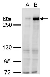
TET1 antibody [N3C1] detects TET1 protein by western blot analysis.
A. 30 μg 293T whole cell lysate/extract
B. 30 μg whole cell lysate/extract of DDDDK-human TET1-transfected 293T cells
5% SDS-PAGE
TET1 antibody [N3C1] (GTX124207) dilution: 1:5000
The HRP-conjugated anti-rabbit IgG antibody (GTX213110-01) was used to detect the primary antibody.
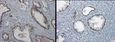
TET1 antibody [N3C1] detects TET1 protein at nucleus on Human normal prostate tissue by immunohistochemical analysis.
Sample: Paraffin-embedded Human normal prostate tissue.
TET1 antibody [N3C1] (GTX124207) dilution: 1:1000.
Antigen Retrieval: Trilogy™ (EDTA based, pH 8.0) buffer, 15min
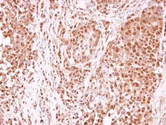
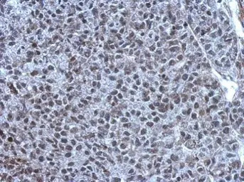
TET1 antibody [N3C1] detects TET1 protein at nucleus in human A549 xenograft by immunohistochemical analysis.
Sample: Paraffin-embedded human A549 xenograft .
TET1 antibody [N3C1] (GTX124207) diluted at 1:250.
Antigen Retrieval: Trilogy™ (EDTA based, pH 8.0) buffer, 15min
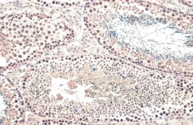
TET1 antibody [N3C1] detects TET1 protein at nucleus by immunohistochemical analysis.
Sample: Paraffin-embedded mouse testis.
TET1 stained by TET1 antibody [N3C1] (GTX124207) diluted at 1:500.
Antigen Retrieval: Citrate buffer, pH 6.0, 15 min
风险提示:丁香通仅作为第三方平台,为商家信息发布提供平台空间。用户咨询产品时请注意保护个人信息及财产安全,合理判断,谨慎选购商品,商家和用户对交易行为负责。对于医疗器械类产品,请先查证核实企业经营资质和医疗器械产品注册证情况。
 文献和实验
文献和实验Application Reference
Chen H et al., Oncogene 2017 (PMID:28346420)
Ciccarone F et al., Oncotarget 2015 (PMID:26136340)
Ichimura N et al., Cancer Prev Res (Phila) 2015 (PMID:26063725)
Chen Y et al., Adv Mater 2016 (PMID:27918113)
Tian Y et al., Scand J Gastroenterol 2016 (PMID:27846738)
Zhang W et al., Sci Rep 2016 (PMID:27708396)
Zhou FC et al., PLoS One 2016 (PMID:27583369)
Yang YA et al., Nucleic Acids Res. 2016 (PMID:27257062)
Ni K et al., Hum Reprod 2016 (PMID:27141042)
Weber AR et al., Nat Commun 2016 (PMID:26932196)
Jin C et al., Nucleic Acids Res 2014 (PMID:24875481)
Jefferson WN et al., Mol Endocrinol 2013 (PMID:24002655)
Dong E et al., Transl Psychiatry 2012 (PMID:22948384)
Hsu CH et al., Cell Rep 2012 (PMID:22999938)
Li J et al., Nat Commun 2022 (PMID:35798741)
Thaler R et al., Nat Commun 2022 (PMID:36202795)
Wu BK et al., Nat Genet 2022 (PMID:35835915)
Kai Qu et al., Int J Biol Sci 2022 (PMID:35342333)
Benedict Shi Xiang Lian et al., iScience 2022 (PMID:35402874)
Shirui Huang et al., Cancer Cell Int 2021 (PMID:33926470)
Jody Ye et al., J Biol Chem 2021 (PMID:33992646)
Lv J et al., Nat Cell Biol 2022 (PMID:35292781)
Feng J et al., Stem Cell Res Ther 2022 (PMID:35073971)
Schagdarsurengin U et al., Clin Epigenetics 2021 (PMID:34844636)
Chu M et al., Front Cell Dev Biol 2021 (PMID:34869387)
Aggarwal RK et al., Proc Natl Acad Sci U S A 2021 (PMID:34551979)
Kawakubo-Yasukochi T et al., Mol Metab 2021 (PMID:34673295)
Chen W et al., Cell Rep 2021 (PMID:34731622)
Patani H et al., Nat Commun 2020 (PMID:32699299)
Huang Z et al., Sci Adv 2021 (PMID:33523915)
Lv H et al., Cell Metab 2021 (PMID:33171124)
Damal Villivalam S et al., Nat Commun 2020 (PMID:32855402)
Gu Y et al., FASEB J 2020 (PMID:32729950)
Rampal R et al., Cell Rep 2014 (PMID:25482556)
Wang J et al., Nutr Metab (Lond) 2020 (PMID:32577122)
Good CR et al., Nucleic Acids Res 2017 (PMID:28531272)
Cao T et al., FASEB J 2020 (PMID:32374060)
Yan YL et al., Front Oncol 2020 (PMID:32528872)
Barazeghi E et al., BMC Cancer 2018 (PMID:30045709)
Cheng Y et al., Cell Rep 2018 (PMID:30540950)
Yu S et al., Int J Endocrinol 2020 (PMID:32089682)
Chen YL et al., Am J Cancer Res 2018 (PMID:30662811)
Xu Y et al., Cell Rep 2020 (PMID:32023451)
Wu J et al., J Exp Clin Cancer Res 2019 (PMID:31399111)
Volnitskiy A et al., PLoS One 2019 (PMID:30730955)
Pei YF et al., Am J Pathol 2019 (PMID:30735628)
Ciccarone F et al., Mol Nutr Food Res 2019 (PMID:30515977)
Zhu J et al., Sci Rep 2017 (PMID:28808304)
Ma F et al., J Cell Physiol 2018 (PMID:30317605)
Fan J et al., Clin Epigenetics 2018 (PMID:30075814)
Ai-hua Wu et al., Current Medical Science 2018 (38)
Atlante S et al., Cell Death Dis 2018 (PMID:29988033)
Adachi K et al., Cell Stem Cell 2018 (PMID:29910149)
Barazeghi E et al., Clin Epigenetics 2016 (PMID:26973719)
Zhou Y et al., Cell Physiol Biochem 2018 (PMID:29558748)
Grosser C et al., Epigenetics 2015 (PMID:26186463)
Ciccarone F et al., Oncotarget 2014 (PMID:24939750)
Tovy A et al., Genes Dev 2017 (PMID:28607180)
Cleven AHG et al., Clin Sarcoma Res 2017 (PMID:28484589)
In the field of therapeutic recombinant proteins, monoclonal antibodies (mAbs) have achieved a rising success with more than 30 mAbs that have reached the market in the past 20 years. From a structural standpoint, one of the most important
Materials 0.1M NaHC03 pH9 DMSO NHS-Biotin (N-Hydroxysuccinimidobiotin, Sigma #H-1759) PBS Procedure Dialyze the sample against carbonate buffer. After dialysis, adjust
Monoclonal Antibody Production Protocol
the rinse twice. Add 100 ul of blocking solution to every well, leave 1 hr at room Temp or O.N at 4°C. PRIMARY ANTIBODY Add the antibody to be tested: Sup of cells = 25 ul, mix well by pipetting up and down (10 times).serum, ascites = 1:100 and a series
 技术资料
技术资料暂无技术资料 索取技术资料

