

![CD68 antibody [ED1]产品图](https://img1.dxycdn.com/2023/0711/451/6106825198874858761-14.jpg)
- ¥6300
- GeneTex

- 美国
- GTX41868
- 2025年07月14日
- WB, ICC/IF, IHC-P, IHC-Fr, FACS, IP, RIA
- Mouse
- Human, Rat, Bovine

企业认证
相关产品推荐更多 >
![LZIP antibody [C1C3]](https://img1.dxycdn.com/2023/0516/045/6925717576889828561-14.jpg!wh200)
![PDGF Receptor alpha antibody [16A1] (APC)](https://img1.dxycdn.com/2023/0711/451/6106825198874858761-14.jpg!wh200)
![RAI3 antibody [N1], N-term](https://img1.dxycdn.com/2023/0711/255/2344116916935858761-14.jpg!wh200)

万千商家帮你免费找货
0 人在求购买到急需产品
相关阅读
香烟烟雾如何加速声带纤维化?CD44-透明质酸轴是关键通路Takara 秋促来袭,提取+反转录+定量套装仅 1200 元abinScience 体内级抗体 全年锁价 低至 ¥1200阿拉丁 22万+ 科研级产品现货速发,品质严控、科研无忧- 详细信息
- 文献和实验
- 技术资料
- 免疫原:
Rat spleen cells
- 亚型:
IgG1
- 形态:
Liquid
- 保存条件:
Store as concentrated solution. Centrifuge briefly prior to opening vial. For short-term storage (1-2 weeks), store at 4ºC. For long-term storage, aliquot and store at -20ºC or below. Avoid multiple freeze-thaw cycles.
- 克隆性:
Monoclonal
- 标记物:
Unconjugated
- 适应物种:
Human, Rat, Bovine
- 保质期:
12 months from the shipping date of the product.
- 抗原来源:
Rat
- 目录编号:
GTX41868
- 级别:
Primary Antibodies
- 库存:
Available
- 供应商:
GeneTex
- 宿主:
Mouse
- 应用范围:
WB, ICC/IF, IHC-P, IHC-Fr, FACS, IP, RIA
- 浓度:
0.5 mg/ml (Please refer to the vial label for the specific concentration.)
- 靶点:
CD68
- 抗体英文名:
CD68 antibody [ED1]
- 抗体名:
CD68 抗体 [ED1]
- 规格:
100 μg

IHC analysis of rat liver, with induced hepatocellular damage tissue using GTX41868 CD68 antibody [ED1].
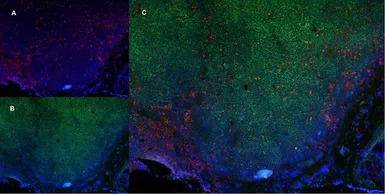
IHC-Fr analysis of rat lymph node tissue using GTX41868 CD68 antibody (red n A) and Mouse anti Rat CD4 (Green in B). C is the merged image with nuclei counter-stained blue using DAPI. Low power.
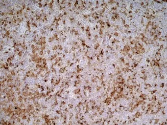
IHC-Fr analysis of rat lymph node tissue using GTX41868 CD68 antibody [ED1].
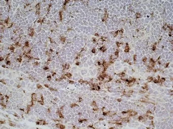
IHC-Fr analysis of rat lymph node tissue using GTX41868 CD68 antibody [ED1].
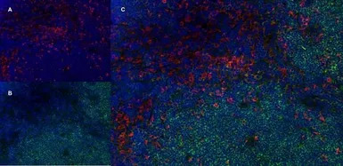
IHC-Fr analysis of rat lymph node tissue using GTX41868 CD68 antibody (red n A) and Mouse anti Rat CD4 (Green in B). C is the merged image with nuclei counter-stained blue using DAPI.Medium power
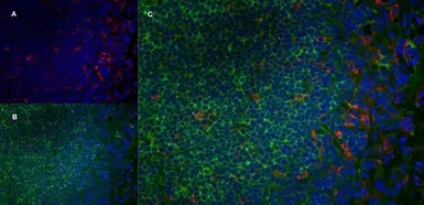
IHC-Fr analysis of rat lymph node tissue using GTX41868 CD68 antibody (red n A) and Mouse anti Rat CD4 (Green in B). C is the merged image with nuclei counter-stained blue using DAPI. High power.
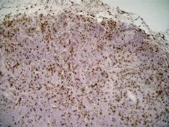
IHC-Fr analysis of rat lymph node tissue using GTX41868 CD68 antibody [ED1].
风险提示:丁香通仅作为第三方平台,为商家信息发布提供平台空间。用户咨询产品时请注意保护个人信息及财产安全,合理判断,谨慎选购商品,商家和用户对交易行为负责。对于医疗器械类产品,请先查证核实企业经营资质和医疗器械产品注册证情况。
 文献和实验
文献和实验Mang Y et al., Ann Transl Med 2022 (PMID:36330418)
Hung HS et al., Cells 2021 (PMID:34831077)
Tseng WC et al., Stem Cell Res Ther 2021 (PMID:34183058)
He S et al., Inflammation 2021 (PMID:33738649)
Kao YJ et al., AJNR Am J Neuroradiol 2019 (PMID:30923084)
Mahara A et al., J Biomed Mater Res A 2020 (PMID:32323458)
Hou HH et al., Am J Physiol Lung Cell Mol Physiol 2018 (PMID:29722565)
Mukwaya A et al., Angiogenesis 2018 (PMID:29445990)
Jeuthe S et al., J Cardiovasc Magn Reson 2016 (PMID:27084492)
Tsutsumi K et al., JPS 2016 (Epub)
Schmerler P et al., Acta Physiol (Oxf) 2014 (PMID:24410878)
Murata K et al., Int J Mol Med 2022 (PMID:35686541)
巨噬细胞 siRNA 递送 “卡脖子”?Absin 助力纳米系统突破降解瓶颈,点亮骨缺损修复新路径
+iNOS+ M1 巨噬细胞,增加 CD68+CD163+ M2 巨噬细胞,营造促修复微环境(图 7,对应原文 Figure 6b)。 图 7 原文 Figure 6b:免疫荧光染色显示 M1(CD68+iNOS+)与 M2(CD68+CD163+)巨噬细胞比例变化 三、Absin 产品助力:关键试剂赋能研究核心环节 该研究的核心机制验证与表型分析中,Absin 的 iNOS 抗体发挥了不可或缺的作用: 产品信息 产品名称 应用场景 iNOS Antibody 免疫
大鼠ED-1/sCD68分子(ED-1)ELISA试剂盒 说明书
上海西唐生物科技有限公司 021-55229872, 65333639 www.westang.com 大鼠 ED-1/sCD68 分子 (ED-1)ELISA 试剂盒 ( 用于血清、血浆、细胞培养上清液和其它生物体液内 ) 原理 本实验采用双抗体夹心 ABC-ELISA 法。用抗大鼠 CD68 单抗包被于酶标板上,标准品和样品中的 CD68 与单抗结合,加入生物素化的抗大鼠 CD68 ,形成免疫
Elabscience|巨噬细胞极化总失败?RAW264.7/THP-1/BMDM/hMDM四大模型避坑指南与实操全攻略
16/32 Antibody,轻轻吹打混匀,室温避光孵育10 min封闭细胞表面的Fc受体; (4)加入各流式抗体(5 μL/test),轻轻吹打混匀,4℃避光孵育30 min; (5)孵育完成后,每管加入1 mL Cell Staining Buffer轻轻吹打混匀,150 × g离心3 min,弃上清; (6)加入200 μL Cell Staining Buffer重悬细胞沉淀,上机检测。 图1. Raw264.7细胞极化培养流程 2. Raw264.7细胞向M1型巨噬细胞极化结果 图
 技术资料
技术资料暂无技术资料 索取技术资料

