


- ¥1700 - 4000
- GeneTex

- 美国
- GTX112760
- 2025年07月15日
- WB, ICC/IF, IHC-P, IP
- Rabbit
- Human, Mouse, Rat

企业认证
相关产品推荐更多 >


万千商家帮你免费找货
0 人在求购买到急需产品
- 详细信息
- 文献和实验
- 技术资料
- 免疫原:
Full length human mtTFA Recombinant protein.
- 亚型:
IgG
- 形态:
Liquid
- 保存条件:
Store as concentrated solution. Centrifuge briefly prior to opening vial. For short-term storage (1-2 weeks), store at 4ºC. For long-term storage, aliquot and store at -20ºC or below. Avoid multiple freeze-thaw cycles.
- 克隆性:
Polyclonal
- 标记物:
Unconjugated
- 适应物种:
Human, Mouse, Rat
- 保质期:
12 months from the shipping date of the product.
- 抗原来源:
Human
- 目录编号:
GTX112760
- 级别:
Primary Antibodies
- 库存:
Available
- 供应商:
GeneTex
- 宿主:
Rabbit
- 应用范围:
WB, ICC/IF, IHC-P, IP
- 浓度:
0.43 mg/ml (Please refer to the vial label for the specific concentration.)
- 靶点:
mtTFA
- 抗体英文名:
mtTFA antibody
- 抗体名:
mtTFA 抗体
- 规格:
100 μl/25 μl
| 规格: | 100 μl | 产品价格: | ¥4000.0 |
|---|---|---|---|
| 规格: | 25 μl | 产品价格: | ¥1700.0 |
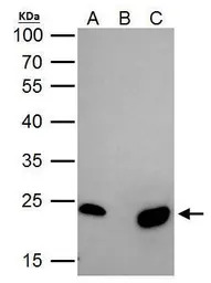
mtTFA antibody immunoprecipitates mtTFA protein in IP experiments. IP Sample: 293T whole cell lysate/extract A. 40 μg 293T whole cell lysate/extract B. Control with 2 μg of preimmune rabbit IgG C. Immunoprecipitation of mtTFA protein by 2 μg of mtTFA antibody (GTX112760) 12% SDS-PAGE The immunoprecipitated mtTFA protein was detected by mtTFA antibody (GTX112760) diluted at 1:1000. EasyBlot anti-rabbit IgG (GTX221666-01) was used as a secondary reagent.

Non-transfected (–) and transfected (+) 293T whole cell extracts (30 μg) were separated by 12% SDS-PAGE, and the membrane was blotted with mtTFA antibody (GTX112760) diluted at 1:3000. The HRP-conjugated anti-rabbit IgG antibody (GTX213110-01) was used to detect the primary antibody.
TFAM antibody detects TFAM protein at mitochondria by immunofluorescent analysis.
Sample: HeLa cells were fixed in 4% PFA at RT for 15 min.
Green: TFAM stained by TFAM antibody (GTX112760) diluted at 1:1000.
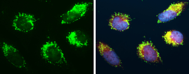
mtTFA antibody detects mtTFA protein at mitochondria by immunofluorescent analysis.
Sample: HeLa cells were fixed in 4% PFA at RT for 15 min.
Green: mtTFA protein stained by mtTFA antibody (GTX112760) diluted at 1:500.
Red: Mitotracker, a mitochondria marker, diluted at 1:2000.
Blue: Hoechst 33342 staining.
Immunohistochemical analysis of paraffin-embedded human breast cancer, using mtTFA(GTX112760) antibody at 1:250 dilution.
Antigen Retrieval: Trilogy™ (EDTA based, pH 8.0) buffer, 15min
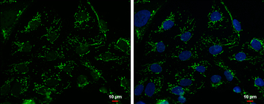
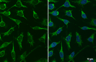
mtTFA antibody detects mtTFA protein at mitochondria by immunofluorescent analysis.
Sample: NT2D1 cells were fixed in 2% PFA/culture medium at RT for 30 min.
Green: mtTFA protein stained by mtTFA antibody (GTX112760) diluted at 1:500.
Blue: Hoechst 33342 staining.
Scale bar = 10 μm.
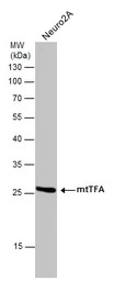
Whole cell extract (30 μg) was separated by 12% SDS-PAGE, and the membrane was blotted with mtTFA antibody (GTX112760) diluted at 1:1000.

Various whole cell extracts (30 μg) were separated by 12% SDS-PAGE, and the membrane was blotted with mtTFA antibody (GTX112760) diluted at 1:500. The HRP-conjugated anti-rabbit IgG antibody (GTX213110-01) was used to detect the primary antibody.
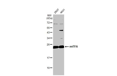
Various whole cell extracts (30 μg) were separated by 12% SDS-PAGE, and the membrane was blotted with mtTFA antibody (GTX112760) diluted at 1:1000. The HRP-conjugated anti-rabbit IgG antibody (GTX213110-01) was used to detect the primary antibody.
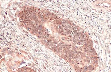
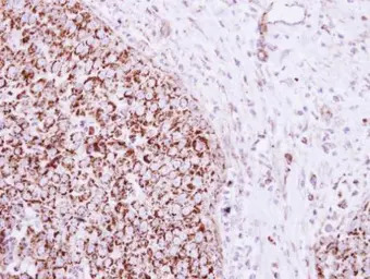
mtTFA antibody detects mtTFA protein at mitochondria by immunohistochemical analysis.
Sample: Paraffin-embedded human esophageal carcinoma.
mtTFA stained by mtTFA antibody (GTX112760) diluted at 1:165.
Antigen Retrieval: Citrate buffer, pH 6.0, 15 min
风险提示:丁香通仅作为第三方平台,为商家信息发布提供平台空间。用户咨询产品时请注意保护个人信息及财产安全,合理判断,谨慎选购商品,商家和用户对交易行为负责。对于医疗器械类产品,请先查证核实企业经营资质和医疗器械产品注册证情况。
 文献和实验
文献和实验Yan H et al., Neuropharmacology 2021 (PMID:34089729)
Yan W et al., Aging (Albany NY) 2021 (PMID:34139672)
Prieto-Carrasco R et al., Biology (Basel) 2021 (PMID:33919054)
Wang Y et al., Aging (Albany NY) 2020 (PMID:33231568)
Qiao A et al., J Cell Biol 2017 (PMID:28183717)
Miao J et al., Aging Cell 2019 (PMID:31318148)
Jinhua Miao et al., Physiol Rep 2021 (PMID:33463897)
Ren B et al., Oxid Med Cell Longev 2022 (PMID:35222795)
Generation of Antibody Molecules Through Antibody Engineering
been overcome to a large extent using genetic-engineering techniques to produce chimeric mouse/human and completely human antibodies. Such an approach is particularly suitable because of the domain structure of the antibody molecule ( 2 ), where functional
The importance of antibody molecules was first recognized in the 1890s, when it was shown that immunity to tetanus and diphtheria was caused by antibodies against the bacterial exotoxins (1 ). Around the same time, it was shown that antisera
General comments: Antibodies, like most proteins, do not like to be freeze-thawed. Avoid repetitive freezing of your solution. The best way to store your antibody is to keep a high protein concentration (>1 mg/ml), add some protease
 技术资料
技术资料暂无技术资料 索取技术资料

