大家都在搜

手机验证

询价列表

暂时没有已询价产品
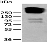
Anti-COA7 antib
ody
企业认证
江西江蓝纯生物试剂有限公司
大量
多克隆
1年
Anti-COA7 antibody
Rabbit
0.9 mg/ml
-20°C
25 μl/100 μl/200 μl

风险提示:丁香通仅作为第三方平台,为商家信息发布提供平台空间。用户咨询产品时请注意保护个人信息及财产安全,合理判断,谨慎选购商品,商家和用户对交易行为负责。对于医疗器械类产品,请先查证核实企业经营资质和医疗器械产品注册证情况。
 技术资料
技术资料
需要更多技术资料 索取更多技术资料
JLC224337.docx 附 (下载 0 次)