


- 询价
- gelatins
- JLC221403
- 上海
- 2025年07月14日
- Rabbit

企业认证
相关产品推荐更多 >


万千商家帮你免费找货
0 人在求购买到急需产品
- 详细信息
- 文献和实验
- 技术资料
- 供应商:
江西江蓝纯生物试剂有限公司
- 库存:
大量
- 克隆性:
多克隆
- 保质期:
1年
- 抗体英文名:
Anti-ATPIF1 antibody
- 宿主:
Rabbit
- 浓度:
0.4 mg/ml
- 保存条件:
-20°C
- 规格:
25 μl/100 μl/200 μl
Cat. No. JLC221403
Package 25 μl/100 μl/200 μl
Storage -20°C, pH7.4 PBS, 0.05% NaN3, 40% Glycerol
Product overview
Description Anti-ATPIF1 rabbit polyclonal antibody
Applications ELISA, WB, IHC
Immunogen Fusion protein of human ATPIF1
Reactivity Human
Content 0.4 mg/ml
Host species Rabbit
Ig class Immunogen-specific rabbit IgG
Purification Antigen affinity purification
Target information
Symbol ATPIF1
Full name ATPase inhibitory factor 1
Synonyms IP; ATPI; ATPIP
Swissprot Q9UII2
Target Background
ATPase inhibitor, mitochondrial is an enzyme that in humans is encoded by the ATPIF1 gene. This gene encodes a mitochondrial ATPase inhibitor. Alternative splicing occurs at this locus and three transcript variants encoding distinct isoforms have been identified. It prevents ATPase from switching to ATP hydrolysis during collapse of the electrochemical gradient, for example during oxygen deprivation ATP synthase inhibitor forms a one to one complex with the F1 ATPase, possibly by binding at the alpha-beta interface. It is thought to inhibit ATP synthesis by preventing the release of ATP. The inhibitor has two oligomeric states, dimer (the active state) and tetramer.
Applications
Immunohistochemistry
Predicted cell location: Cytoplasm
Positive control: Human thyroid cancer
Recommended dilution: 50-200 Predicted cell location: Cytoplasm
Positive control: Human breast cancer
Recommended dilution: 50-200
The image on the left is immunohistochemistry of paraffin-embedded Human thyroid cancer tissue using ml221403(ATPIF1 Antibody) at dilution 1/60, on the right is treated with fusion protein. (Original magnification: ×200) The image on the left is immunohistochemistry of paraffin-embedded Human breast cancer tissue using ml221403(ATPIF1 Antibody) at dilution 1/60, on the right is treated with fusion protein. (Original magnification: ×200)
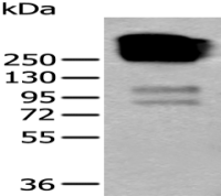
Western blotting
Predicted band size:12 kDa
Positive control:Hela, Jurkat, MCF7 and A431 cells
Recommended dilution: 1000-5000
Gel: 10+12+15%SDS-PAGE
Lysate: 40 μg
Lane 1-4: Hela cells, Jurkat cells, MCF7 cells, A431 cells
Primary antibody: ml221403(ATPIF1 Antibody) at dilution 1/1350
Secondary antibody: Goat anti rabbit IgG at 1/8000 dilution
Exposure time: 1 minute
ELISA
Recommended dilution: 2000-10000
风险提示:丁香通仅作为第三方平台,为商家信息发布提供平台空间。用户咨询产品时请注意保护个人信息及财产安全,合理判断,谨慎选购商品,商家和用户对交易行为负责。对于医疗器械类产品,请先查证核实企业经营资质和医疗器械产品注册证情况。
 文献和实验
文献和实验a simple ELISA method to detect human anti-antibody response.
Purification Of Anti Peptide Antibody
antibody that could contribute to staining background. The proportion in each pool varies with the peptide immunogen. The quality of the anti-peptide serum seems to increase with multiple boosts - the first bleed may be feeble.
Anticardiolipin Antibody and Anti-beta 2 Glycoprotein I Antibody Assays
standard” laboratory test to diagnose or classify a patient as having APS. This chapter discusses the clinical and laboratory theoretical and technical aspects of aCL and anti-β2GPI antibody assays.


