

- ¥1800
- 中乔新舟

- 中国
- ZQ0904
- 2025年12月18日

企业认证

万千商家帮你免费找货
0 人在求购买到急需产品
- 详细信息
- 文献和实验
- 技术资料
- 英文名:
Y79
- 库存:
大量
- 供应商:
中乔新舟
- 细胞类型:
细胞系
- 品系:
视觉系统
- 组织来源:
视觉系统
- 相关疾病:
否
- 物种来源:
视觉系统
- 免疫类型:
否
- 细胞形态:
咨询销售
- 器官来源:
视觉系统
- 运输方式:
T25瓶运输
- 年限:
5-10年
- 生长状态:
贴壁生长
- 规格:
5 x 10^5 cells/vial
|
产品名称 |
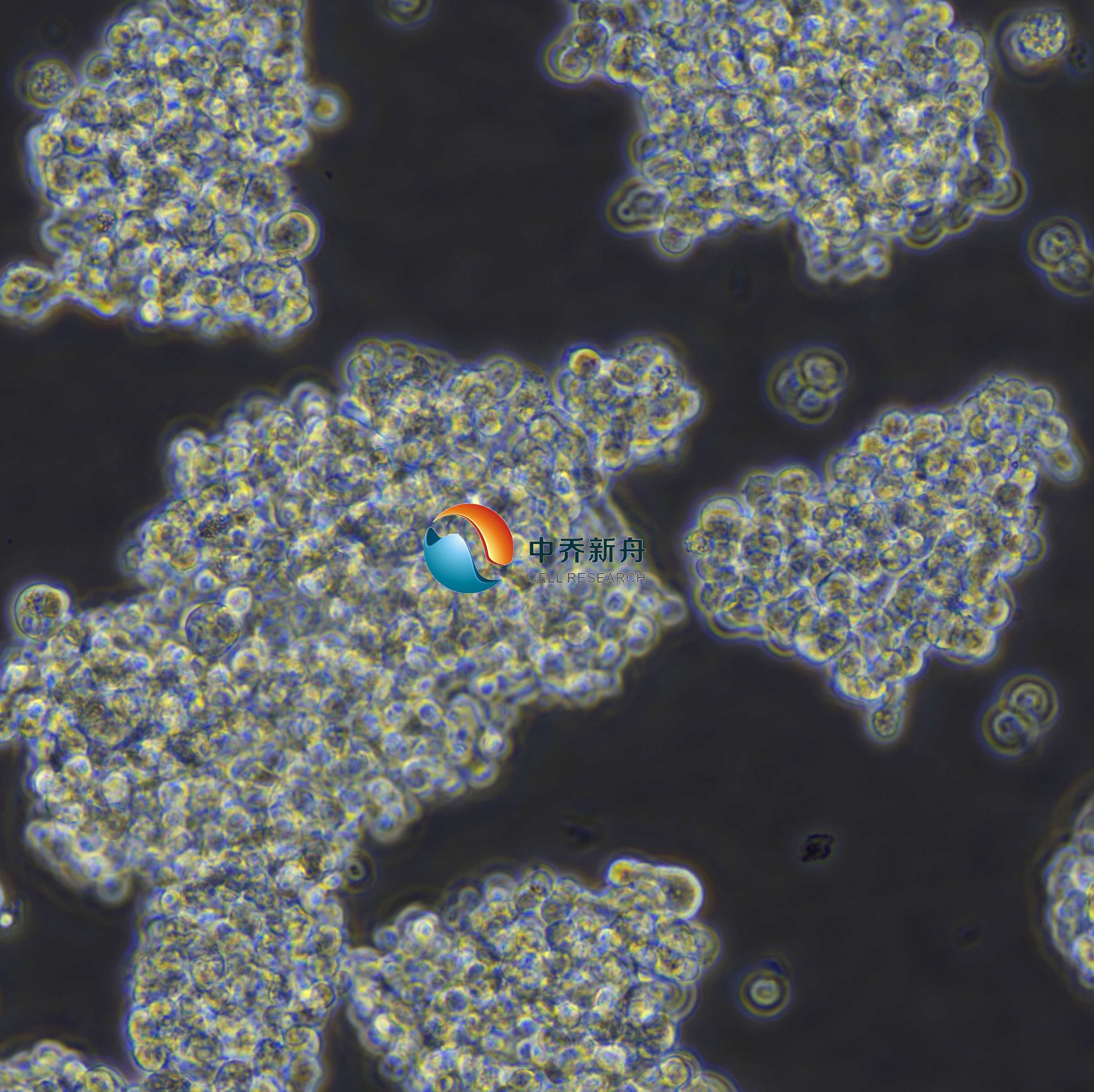
Y79人视网膜母细胞瘤细胞 |
|
货号 |
ZQ0904 |
|
产品介绍 |
这株y79系由t.w. reid及其同事于1971年1月通过摘除后立即获得的右眼原发肿瘤的外植体培养分离。捐赠者有很强的视网膜母细胞瘤家族史。据报道,超微结构特征包括核膜信息、三重膜结构、微管、大包膜泡、中心粒、基体和环状片层与原肿瘤相似。 注意事项: Y79细胞悬浮聚团生长,细胞传代时需要冲散细胞为单细胞后传代。细胞复苏生长较慢,需要1周左右生长。 |
|
种属 |
人 |
|
性别/年龄 |
女/2.5岁 |
|
组织 |
眼睛,视网膜 |
|
疾病 |
视网膜母细胞瘤;视网膜神经胶质细胞瘤 |
|
细胞类型 |
肿瘤细胞 |
|
形态学 |
多细胞集群、呈葡萄状聚集 |
|
生长方式 |
悬浮 |
|
倍增时间 |
大约1周左右 |
|
培养基和添加剂 |
RPMI-1640(中乔新舟 货号:ZQ-200)+20%胎牛血清(中乔新舟 货号:ZQ0500)+1%P/S(中乔新舟 货号:CSP006) |
|
推荐完全培养基货号 |
ZM0904 |
|
生物安全等级 |
BSL-1 |
|
STR位点信息 |
Amelogenin: X CSF1PO: 11,12 |
|
培养条件 |
95%空气,5%二氧化碳;37℃ |
|
抗原表达/受体表达 |
*** |
|
基因表达 |
*** |
|
保藏机构 |
ATCC; HTB-18 BCRC; 60422 BCRJ; 0380 DSMZ; ACC-246 ECACC; 86093003 |
|
供应限制 |
仅供科研使用 |
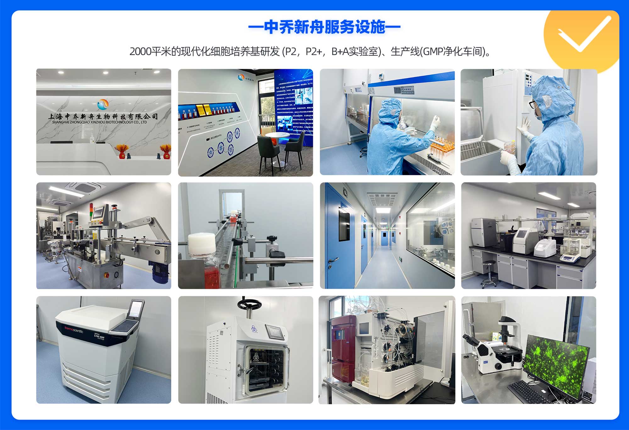
上海中乔新舟生物科技有限公司成立于2011年,历经十多年发展,主要专注于细胞生物学产品的研究和开发,专注于为药企、各类科研机构及CRO企业提供符合标准规范的细胞培养服务、细胞培养基、细胞检测试剂盒、细胞培养试剂,胎牛血清和细胞生物学技术服务等。
公司一直致力于为高等院校、研究机构、医院、CRO及CDMO企业提供细胞培养完整解决方案,这些产品旨在满足细胞培养的多样需求,确保实验和研究的有效进行。引用中乔新舟(ZQXZBIO)产品和服务的文献超数千篇。
产品服务
细胞资源:原代细胞、细胞株、干细胞、示踪细胞、耐药株细胞、永生化细胞等基因工程细胞。
试剂产品:胎牛血清、完全培养基(适用于原代细胞及细胞株)、无血清培养基、基础培养基、细胞转染试剂、重组因子、胰酶和双抗等等细胞培养所有实验相关产品。
技术服务:稳转株构建、原代细胞分离、特殊培养基定制服务、细胞检测等。

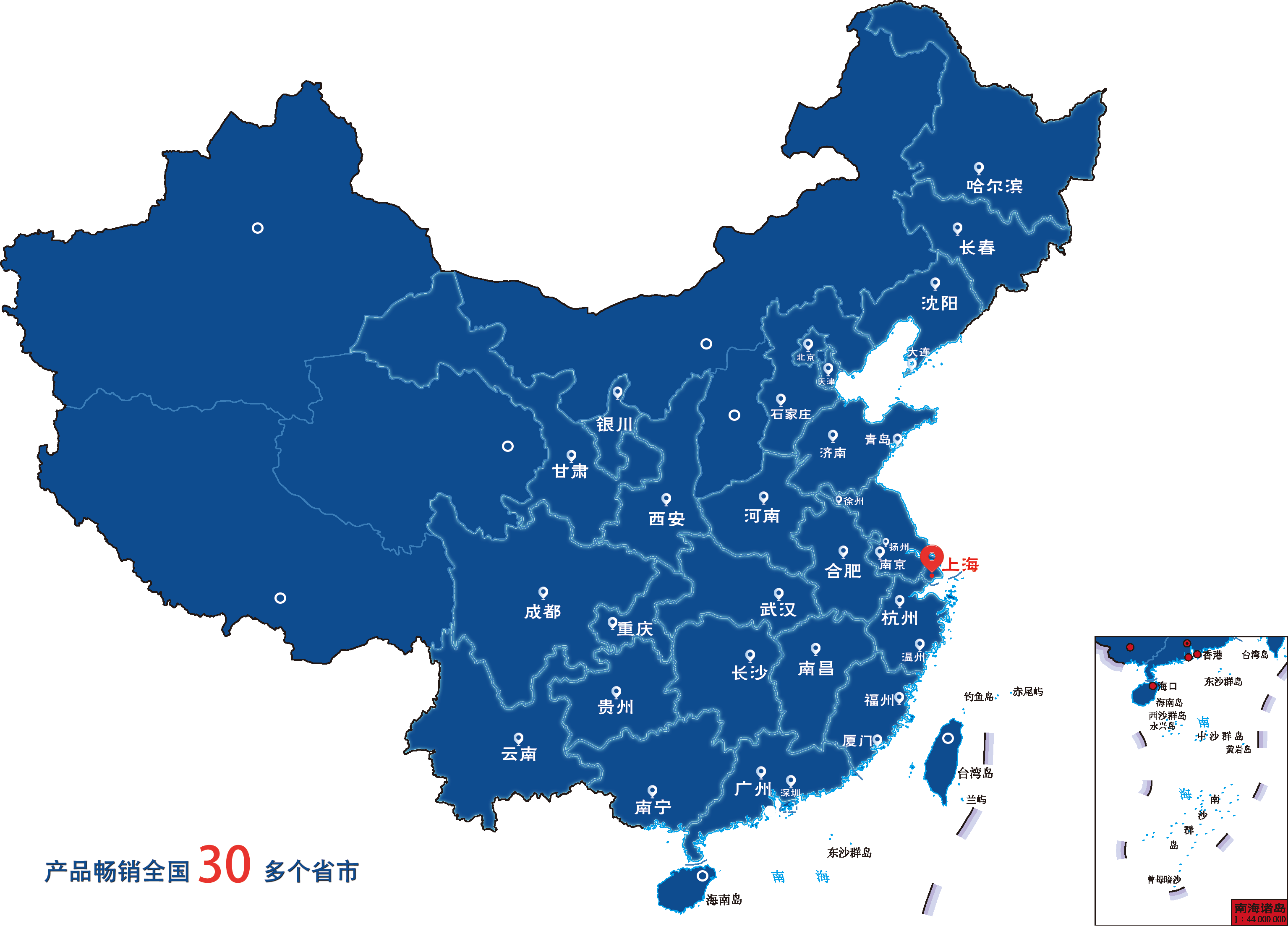
目前产品已经畅销国内30多个省市,与客户建立长期的合作伙伴关系,共同实现成功。全体员工将不懈努力,继续为科研人员提供优良的产品和服务,致力成为全球细胞培养领域的参与者。
企业愿景
致力于成为国内细胞培养基产业的佼佼者,生物医药领域上游原材料的优良提供商。
企业使命
成长为专业细胞系及原代细胞培养供应商、专业细胞培养基及培养试剂生产商。
企业荣誉


风险提示:丁香通仅作为第三方平台,为商家信息发布提供平台空间。用户咨询产品时请注意保护个人信息及财产安全,合理判断,谨慎选购商品,商家和用户对交易行为负责。对于医疗器械类产品,请先查证核实企业经营资质和医疗器械产品注册证情况。
 文献和实验
文献和实验论文标题: Downregulation of miR-211-5p Promotes Carboplatin Resistance in Human Retinoblastoma Y79 Cells by Affecting the GDNF–LIF Interaction
DOI: 10.3389/fonc.2022.848733
发表时间: 2022-03-02
期刊: Frontiers in Oncology
影响因子: 6.244
货号: ZQ0904
产品名称: human retinoblastoma cell line Y79
PubMed=4135597; DOI=10.1093/jnci/53.2.347
Reid T.W., Albert D.M., Rabson A.S., Russell P., Craft J.L., Chu E.W., Tralka T.S., Wilcox J.L.
Characteristics of an established cell line of retinoblastoma.
J. Natl. Cancer Inst. 53:347-360(1974)
PubMed=679190
McFall R.C., Nagy R.M., Nagle B.T., McGreevy L.M.
Scanning electron microscopic observation of two retinoblastoma cell lines.
Cancer Res. 38:2827-2835(1978)
PubMed=6167750; DOI=10.1093/jnci/67.2.301
Gilbert F., Balaban G.B., Breg W.R., Gallie B.L., Reid T.W., Nichols W.W.
Homogeneously staining region in a retinoblastoma cell line: relevance to tumor initiation and progression.
J. Natl. Cancer Inst. 67:301-306(1981)
PubMed=6351622; DOI=10.1016/S0002-9394(14)77822-5
Ohashi Y., Sasabe T., Nishida T., Nishi Y., Higashi H.
Hanganutziu-Deicher heterophile antigen in human retinoblastoma cells.
Am. J. Ophthalmol. 96:321-325(1983)
DOI=10.5795/jjscc.24.451
Tsumuraya M., Nakajima T., Terasaki T., Kodama T., Shimosato Y., Higuchi H., Uei Y.
Analysis of LDH isoenzyme patterns in cell lines of the small cell carcinoma (lung) and retinoblastoma.
Nihon Rinsho Saibo Gakkai Zasshi 24:451-456(1985)
PubMed=3518877; DOI=10.3109/07357908609038260
Fogh J.
Human tumor lines for cancer research.
Cancer Invest. 4:157-184(1986)
PubMed=3413073; DOI=10.1073/pnas.85.16.6017
Lee E.Y.-H.P., Bookstein R., Young L.-J., Lin C.-J., Rosenfeld M.G., Lee W.-H.
Molecular mechanism of retinoblastoma gene inactivation in retinoblastoma cell line Y79.
Proc. Natl. Acad. Sci. U.S.A. 85:6017-6021(1988)
PubMed=2917337; DOI=10.1016/0165-4608(89)90079-4
Horsthemke B., Greger V., Becher R., Passarge E.
Mechanism of i(6p) formation in retinoblastoma tumor cells.
Cancer Genet. Cytogenet. 37:95-102(1989)
PubMed=1679230; DOI=10.3109/13816819109023085
Madreperla S.A., Bookstein R., Jones O.W., Lee W.-H.
Retinoblastoma cell lines Y79, RB355 and WERI-Rb27 are genetically related.
Ophthalmic Paediatr. Genet. 12:49-56(1991)
CLPUB00447
Mulivor R.A., Suchy S.F.
1992/1993 catalog of cell lines. NIGMS human genetic mutant cell repository. 16th edition. October 1992.
(In) Institute for Medical Research (Camden, N.J.) NIH 92-2011; pp.1-918; National Institutes of Health; Bethesda (1992)
PubMed=7689221; DOI=10.1073/pnas.90.16.7578
Godbout R., Squire J.A.
Amplification of a DEAD box protein gene in retinoblastoma cell lines.
Proc. Natl. Acad. Sci. U.S.A. 90:7578-7582(1993)
PubMed=7499444; DOI=10.1007/BF01213319
Inomata M., Saijo N., Kawashima K., Kaneko A., Fujiwara Y., Kunikane H., Tanaka Y.
Induction of apoptosis in cultured retinoblastoma cells by the protein phosphatase inhibitor, okadaic acid.
J. Cancer Res. Clin. Oncol. 121:729-738(1995)
PubMed=18036396; DOI=10.1016/j.cancergencyto.2007.08.014
Paderova J., Orlic-Milacic M., Yoshimoto M., da Cunha Santos G., Gallie B.L., Squire J.A.
Novel 6p rearrangements and recurrent translocation breakpoints in retinoblastoma cell lines identified by spectral karyotyping and mBAND analyses.
Cancer Genet. Cytogenet. 179:102-111(2007)
PubMed=18799932; DOI=10.1097/MPH.0b013e31816e232d
Kim J.H., Kim J.H., Yu Y.S., Kim D.H., Kim Y.K., Kim K.-W.
Comparative genomic hybridization analysis of newly established retinoblastoma cell lines of adherent growth compared with Y79 of nonadherent growth.
J. Pediatr. Hematol. Oncol. 30:571-574(2008)
PubMed=19686387; DOI=10.1111/j.1471-4159.2009.06322.x
Glubrecht D.D., Kim J.-H., Russell L., Bamforth J.S., Godbout R.
Differential CRX and OTX2 expression in human retina and retinoblastoma.
J. Neurochem. 111:250-263(2009)
PubMed=20215515; DOI=10.1158/0008-5472.CAN-09-3458
Rothenberg S.M., Mohapatra G., Rivera M.N., Winokur D., Greninger P., Nitta M., Sadow P.M., Sooriyakumar G., Brannigan B.W., Ulman M.J., Perera R.M., Wang R., Tam A., Ma X.-J., Erlander M., Sgroi D.C., Rocco J.W., Lingen M.W., Cohen E.E.W., Louis D.N., Settleman J., Haber D.A.
A genome-wide screen for microdeletions reveals disruption of polarity complex genes in diverse human cancers.
Cancer Res. 70:2158-2164(2010)
PubMed=21697133; DOI=10.1167/iovs.11-7479
Oshikawa M., Tsutsui C., Ikegami T., Fuchida Y., Matsubara M., Toyama S., Usami R., Ohtoko K., Kato S.
Full-length transcriptome analysis of human retina-derived cell lines ARPE-19 and Y79 using the vector-capping method.
Invest. Ophthalmol. Vis. Sci. 52:6662-6670(2011)
PubMed=23498719; DOI=10.1016/S1470-2045(13)70045-7
Rushlow D.E., Mol B.M., Kennett J.Y., Yee S., Pajovic S., Theriault B.L., Prigoda-Lee N.L., Spencer C., Dimaras H., Corson T.W., Pang R., Massey C., Godbout R., Jiang Z., Zacksenhaus E., Paton K., Moll A.C., Houdayer C., Raizis A., Halliday W., Lam W.L., Boutros P.C., Lohmann D.R., Dorsman J.C., Gallie B.L.
Characterisation of retinoblastomas without RB1 mutations: genomic, gene expression, and clinical studies.
Lancet Oncol. 14:327-334(2013)
视网膜母细胞瘤是起源于视网膜的胚胎性的眼内恶性肿瘤。本病与遗传、染色体畸变、病毒感染有关。患者90%为3岁以内婴儿,少数发生于大龄儿童,偶见成人。无种族及性别差异。多单眼发病,约占70%,双眼同时或先后发病者约为30%。双眼发病者全部及单侧患者中的10—15%多具有遗传性,属常染色体显性遗传,外显率不全,故表面正常,带有致病基因的父母不一定发病,但可以有患病的子女。在我国有家族史者占2.5%—3.5%。染色体研究也证明部分患者有染色体的异常,发现13号染色体长臂缺失或基因
肿瘤的出现:比如双侧乳腺癌,双侧肾癌,双侧视网膜母细胞瘤。 多个原发性肿瘤在同一个病人身上出现:如一个患者既有原发性肠癌,又有原发性卵巢癌。 非肿瘤临床表现的出现:如Cowden综合征,该疾病是一种罕见的常染色体显性遗传病,主要表现为多重错构瘤及某类肿瘤发生风险的增加。临床表现多种多样,包括乳腺癌、子宫内膜癌、甲状腺癌等,皮肤方面表现为口腔及皮肤乳头状瘤,胃肠方面表现为混合性息肉(包括错构瘤)。 无外环境风险因素的发现:一般我们认为肿瘤是复杂疾病,肿瘤的发生是遗传因素与环境因素(包括
与原癌基因编码的蛋白质促进细胞生长相反,在正常情况下存在于细胞内的另一类基因——肿瘤抑制基因的产物能抑制细胞的生长。若其功能丧失则可能促进细胞的肿瘤性转化。由此看来,肿瘤的发生可能是癌基因的激活与肿瘤抑制基因的失活共同作用的结果。目前了解最多的两种肿瘤抑制基因是Rb基因和P53基因。它们的产物都是以转录调节因子的方式控制细胞生长的核蛋白。其它肿瘤抑制基因还有神经纤维瘤病-1基因、结肠腺瘤性息肉基因、结肠癌丢失基因和Wilms瘤-1等。 Rb基因随着对一种少见的儿童肿瘤——视网膜母细胞瘤
 技术资料
技术资料暂无技术资料 索取技术资料

