


- ¥2980
- LMAI Bio
- LM-11788R-FITC
- 中国/美国/欧洲
- 2026年05月29日
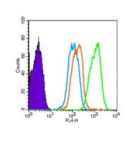
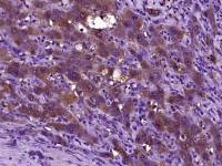
- ICC=1:50-200 IF=1:50-200
- Human, Mouse, Rat, Cow, Horse,
- Human, Mouse, Rat, Cow, Horse,

企业认证
相关产品推荐更多 >


万千商家帮你免费找货
0 人在求购买到急需产品
- 详细信息
- 询价记录
- 文献和实验
- 技术资料
- 供应商:
上海联迈生物工程有限公司
- 库存:
大量
- 靶点:
详见说明书
- 级别:
1
- 目录编号:
LM-11788R-FITC
- 克隆性:
多克隆
- 抗原来源:
Rabbit
- 保质期:
1年
- 抗体英文名:
Anti-Prion protein PrP/CD230/FITC
- 抗体名:
Anti-Prion protein PrP/CD230/FITC
- 标记物:
FITC标记
- 宿主:
Human, Mouse, Rat, Cow, Horse,
- 适应物种:
Human, Mouse, Rat, Cow, Horse,
- 免疫原:
详见说明书
- 亚型:
IGg
- 形态:
粉末、液体、冻干粉
- 应用范围:
ICC=1:50-200 IF=1:50-200
- 浓度:
1mg/ml
- 保存条件:
-20 °C
- 规格:
100ul
| 英文名称 | Anti-Prion protein PrP/CD230/FITC |
| 中文名称 | FITC标记的朊病毒蛋白CD230抗体 |
| 别 名 | AltPrP; ASCR; atal familial insomnia; CD230; CD230 antigen; CJD; Creutzfeld Jakob disease; Gerstmann-Strausler-Scheinker syndrome; GSS; KURU; Major prion protein; PRIO_HUMAN. |
| 规格价格 | 100ul/2980元 购买 大包装/询价 |
| 说 明 书 | 100ul |
| 研究领域 | 细胞生物 神经生物学 干细胞 细菌及病毒 细胞表面分子 |
| 抗体来源 | Rabbit |
| 克隆类型 | Polyclonal |
| 交叉反应 | Human, Mouse, Rat, Cow, Horse, |
| 产品应用 | ICC=1:50-200 IF=1:50-200 not yet tested in other applications. optimal dilutions/concentrations should be determined by the end user. |
| 分 子 量 | 25kDa |
| 细胞定位 | 细胞膜 |
| 性 状 | Lyophilized or Liquid |
| 浓 度 | 1mg/ml |
| 免 疫 原 | KLH conjugated synthetic peptide derived from human Prion protein PrP/CD230 |
| 亚 型 | IgG |
| 纯化方法 | affinity purified by Protein A |
| 储 存 液 | 0.01M TBS(pH7.4) with 1% BSA, 0.03% Proclin300 and 50% Glycerol. |
| 保存条件 | Store at -20 °C for one year. Avoid repeated freeze/thaw cycles. The lyophilized antibody is stable at room temperature for at least one month and for greater than a year when kept at -20°C. When reconstituted in sterile pH 7.4 0.01M PBS or diluent of antibody the antibody is stable for at least two weeks at 2-4 °C. |
| 产品介绍 | background: The function of PrP is still under debate. May play a role in neuronal development and synaptic plasticity. May be required for neuronal myelin sheath maintenance. May play a role in iron uptake and iron homeostasis (By similarity). Isoform 2 may act as a growth suppressor by arresting the cell cycle at the G0/G1 phase. Soluble oligomers are toxic to cultured neuroblastoma cells and induce apoptosis (in vitro). Function: The function of PrP is still under debate. May play a role in neuronal development and synaptic plasticity. May be required for neuronal myelin sheath maintenance. May play a role in iron uptake and iron homeostasis (By similarity). Isoform 2 may act as a growth suppressor by arresting the cell cycle at the G0/G1 phase. Soluble oligomers are toxic to cultured neuroblastoma cells and induce apoptosis (in vitro). Subunit: Monomer and homodimer. Has a tendency to aggregate into amyloid fibrils containing a cross-beta spine, formed by a steric zipper of superposed beta-strands. Soluble oligomers may represent an intermediate stage on the path to fibril formation. Copper binding may promote oligomerization. Interacts with GRB2, APP, ERI3/PRNPIP and SYN1. Mislocalized cytosolically exposed PrP interacts with MGRN1; this interaction alters MGRN1 subcellular location and causes lysosomal enlargement (By similarity). Interacts with KIAA1191. Subcellular Location: Cell membrane. Golgi apparatus and Cytoplasm. Nucleus. Accumulates outside the secretory route in the cytoplasm, from where it relocates to the nucleus. Post-translational modifications: The glycosylation pattern (the amount of mono-, di- and non-glycosylated forms or glycoforms) seems to differ in normal and CJD prion. Isoform 2 is sumoylated by SUMO1. DISEASE: Note=PrP is found in high quantity in the brain of humans and animals infected with neurodegenerative diseases known as transmissible spongiform encephalopathies or prion diseases, like: Creutzfeldt-Jakob disease (CJD), fatal familial insomnia (FFI), Gerstmann-Straussler disease (GSD), Huntington disease-like type 1 (HDL1) and kuru in humans; scrapie in sheep and goat; bovine spongiform encephalopathy (BSE) in cattle; transmissible mink encephalopathy (TME); chronic wasting disease (CWD) of mule deer and elk; feline spongiform encephalopathy (FSE) in cats and exotic ungulate encephalopathy (EUE) in nyala and greater kudu. The prion diseases illustrate three manifestations of CNS degeneration: (1) infectious (2) sporadic and (3) dominantly inherited forms. TME, CWD, BSE, FSE, EUE are all thought to occur after consumption of prion-infected foodstuffs. Defects in PRNP are the cause of Creutzfeldt-Jakob disease (CJD) [MIM:123400]. CJD occurs primarily as a sporadic disorder (1 per million), while 10-15% are familial. Accidental transmission of CJD to humans appears to be iatrogenic (contaminated human growth hormone (HGH), corneal transplantation, electroencephalographic electrode implantation, etc.). Epidemiologic studies have failed to implicate the ingestion of infected annimal meat in the pathogenesis of CJD in human. The triad of microscopic features that characterize the prion diseases consists of (1) spongiform degeneration of neurons, (2) severe astrocytic gliosis that often appears to be out of proportion to the degree of nerve cell loss, and (3) amyloid plaque formation. CJD is characterized by progressive dementia and myoclonic seizures, affecting adults in mid-life. Some patients present sleep disorders, abnormalities of high cortical function, cerebellar and corticospinal disturbances. The disease ends in death after a 3-12 months illness. Defects in PRNP are the cause of fatal familial insomnia (FFI) [MIM:600072]. FFI is an autosomal dominant disorder and is characterized by neuronal degeneration limited to selected thalamic nuclei and progressive insomnia. Defects in PRNP are the cause of Gerstmann-Straussler disease (GSD) [MIM:137440]. GSD is a heterogeneous disorder and was defined as a spinocerebellar ataxia with dementia and plaquelike deposits. GSD incidence is less than 2 per 100 million live births. Defects in PRNP are the cause of Huntington disease-like type 1 (HDL1) [MIM:603218]. HDL1 is an autosomal dominant, early onset neurodegenerative disorder with prominent psychiatric features. Defects in PRNP are the cause of kuru (KURU) [MIM:245300]. Kuru is transmitted during ritualistic cannibalism, among natives of the New Guinea highlands. Patients exhibit various movement disorders like cerebellar abnormalities, rigidity of the limbs, and clonus. Emotional lability is present, and dementia is conspicuously absent. Death usually occurs from 3 to 12 month after onset. Defects in PRNP are the cause of spongiform encephalopathy with neuropsychiatric features (SENF) [MIM:606688]; an autosomal dominant presenile dementia with a rapidly progressive and protracted clinical course. The dementia was characterized clinically by frontotemporal features, including early personality changes. Some patients had memory loss, several showed aggressiveness, hyperorality and verbal stereotypy, others had parkinsonian symptoms. Prion diseases, or transmissible spongiform encephalopathies (TSEs), are manifested as genetic, infectious or sporadic, lethal neurodegenerative disorders involving alterations of the prion protein (PrP). Characteristic of prion diseases, cellular PrP (PrPc) is converted to the disease form, PrPSc, through alterations in the protein folding conformations. PrPc is constitutively expressed in normal adult brain and is sensitive to proteinase K digestion, while the altered PrPSc conformation is resistant to proteases, resulting in a distinct molecular mass after PK treatment. Consistent with the transient infection process of prion diseases, incubation of PrPc with PrPSc both in vitro and in vivo produces PrPc that is resistant to protease degradation. Infectious PrPSc is found at high levels in the brains of animals affected by TSEs, including scrapie in sheep, BSE in cattle and Cruetzfeldt-Jakob disease in humans. Similarity: Belongs to the prion family. Database links: Entrez Gene: 281427 Cow Entrez Gene: 5621 Human Entrez Gene: 19122 Mouse Entrez Gene: 24686 Rat Entrez Gene: 493887 Sheep Omim: 176640 Human SwissProt: P10279 Cow SwissProt: P04156 Human SwissProt: P04925 Mouse SwissProt: P13852 Rat SwissProt: P23907 Sheep Unigene: 472010 Human Unigene: 610285 Human Unigene: 727471 Human Unigene: 648 Mouse Unigene: 3936 Rat Important Note: This product as supplied is intended for research use only, not for use in human, therapeutic or diagnostic applications. |
风险提示:丁香通仅作为第三方平台,为商家信息发布提供平台空间。用户咨询产品时请注意保护个人信息及财产安全,合理判断,谨慎选购商品,商家和用户对交易行为负责。对于医疗器械类产品,请先查证核实企业经营资质和医疗器械产品注册证情况。
- 作者
- 内容
- 询问日期
 文献和实验
文献和实验接收范围。 一般来说,实验panel有几种荧光,就需要设置几个单阳管。 同型对照(Isotype Control),是指与使用的流式抗体具有相同种属来源、亚型、荧光标记物、使用剂量和浓度,但对目标靶点无特异性结合的抗体。同型对照通常用来消除抗体的非特异性结合造成的背景染色。 FMO 对照是 Fluorescence Minus One(荧光扣除一)的缩写。在多色实验中,可能会出现某些通道信号难以区分的情况,主要原因是其他某个荧光对该通道的干扰很大,不能使用未染色的空白对照或全部用同型对照来设门
多色流式实验中,由于不同荧光素的发射波长的覆盖范围不同,可能产生重叠,对实验结果的数据分析造成一定干扰,我们通常会设置单阳管进行调节补偿。同时,单阳管还可以辅助我们调节通道电压,防止信号超出接收范围。 图示:常见荧光素的发射光谱 A. 如何设置单阳管 单阳管即单染管,是只添加一种荧光抗体的样本管。一般来说,实验 panel 有几种荧光,就需要设置几个单阳管。如:APC、PE、FITC 的三色 panel,需要设置三个单阳管。 B. 制备单阳管的要求 ①单阳管和全染管的样本类型、荧光素要保持
内腔可产生高强度的磁场,足以分离磁微粒标记的细胞,无需分离柱提供提供更高强度的磁场。由于磁微粒极小,不会影响靶细胞的流式分析结果,因此不需要去除。除了现有的定型产品外,客户可选择EasySep® PE、FITC或生物素分选系统与自己的PE、FITC或生物素—偶联的单抗相结合,来富集任何所需的细胞。如果您有自己的小鼠IgG1抗任何细胞的抗体, 也可以使用“Do-It-Yourself”试剂盒做成TAC,用来分选您想要的细胞。此外,还可以根据您的特殊需要,专门为您设计定做试剂盒。用于正选小鼠的细胞标记
 技术资料
技术资料暂无技术资料 索取技术资料

