产品封面图

Flag Tag标签抗体

收藏
  • ¥1250
  • AFFINTY
  • T0003
  • USA
  • 2025年11月14日
  • IHC/WB/ELISA
  • Mouse
  • all
avatar
  • 企业认证

    点击 QQ 联系

    • 详细信息
    • 文献和实验
    • 技术资料
    • 抗体名

      Flag Tag标签抗体

    • 抗体英文名

      Flag-Tag Antibody - #T0003

    • 抗原

      Flag Tag标签抗体

    • 浓度

      Flag-Tag Antibody - #T0003

    • 应用范围

      IHC/WB/ELISA

    • 宿主

      Mouse

    • 适应物种

      all

    • 库存

      T0003

    • 目录编号

      T0003

    • 标记物

      Flag Tag标签抗体

    • 保存条件

      低温

    • 规格

      50ul

    Flag Tag标签抗体
    Flag-Tag Antibody - #T0003
    产品: Flag-Tag 抗体
    货号: T0003
    来源: Mouse
    应用: WB, IHC, IF/ICC, IP, ELISA
    反应: All
    蛋白号:
    RRID: AB_2839412
    来源:
    Mouse
    应用:
    WB 1:3000-1:10000, IF/ICC: 1:200, IP 1:200, IHC 1:50-1:200
    *The optimal dilutions should be determined by the end user.
    反应:
    All
    克隆:
    Monoclonal [T177]
    RRID:
    AB_2839412
    引用格式: Affinity Biosciences Cat# T0003, RRID:AB_2839412.
    纯化:
    Affinity-chromatography.
    保存:
    Mouse IgG1 in phosphate buffered saline (without Mg2+ and Ca2+), pH 7.4, 150mM NaCl, 0.02% sodium azide and 50% glycerol. Store at -20 °C. Stable for 12 months from date of receipt.
    别名:

    展开/折叠

    DDDDK epitope tag; DDDK; ddk; DYKDDDDK; DYKDDDDK epitope tag; DYKDDDDK tag; ECS epitope tag; ECS tag; Enterokinase Cleavage Site epitope tag; Enterokinase Cleavage Site tag; FLAG; FLAG tag antibody

    1). Liu H et al. Psychologic Stress Drives Progression of Malignant Tumors via DRD2/HIF1α Signaling. Cancer Res 2021 Oct 15;81(20):5353-5365. (PubMed: 34321238) [IF=13.312]
    产品细节图片1

    Application: WB Species: mouse Sample:

    Fig. 4 |DRD2 overexpression inhibited the ubiquitin-dependent degradation of HIF1α. N, Effect of DRD2 on the content of ubiquitin binding to HIF1α. DRD2-overexpressing cells treated with MG132, and cellular extracts were prepared for co-IP assay with anti–HIF1α antibody, followed by Western blotting detection of ubiquitin. siRNA, small interfering RNA. ns, nonsignificant; *, P < 0.05; **, P < 0.01

    2). Liu W et al. Micheliolide Ameliorates Diabetic Kidney Disease by Inhibiting Mtdh-mediated Renal Inflammation in Type 2 Diabetic db/db Mice. Pharmacol Res 2019 Oct 24:104506 (PubMed: 31669149) [IF=10.334]

    3). Ye F et al. circFBXW7 Inhibits Malignant Progression by Sponging miR-197-3p and Encoding a 185-aa Protein in Triple-Negative Breast Cancer. Mol Ther Nucleic Acids 2019 Aug 14;18:88-98 (PubMed: 31536884) [IF=10.183]
    产品细节图片2

    Application: WB Species: human Sample: BT549 cells

    Figure 6.| FBXW7-185aa, Encoded by circFBXW7, Inhibited the Proliferation and Metastasis of TNBC Cells through Downregulating c-Myc Expression.s. (H)FBXW7-185aa-FLAG-overexpressing BT549 cells were cotransfected with sh-FBXW7 or USP28 vectors. The FBXW7-185aaFLAG, FBXW7, USP28, and c-Myc expression levels were quantified by western blot analysis.

    4). Matsuoka TA et al. Mafa Enables Pdx1 to Effectively Convert Pancreatic Islet Progenitors and Committed Islet α-Cells Into β-Cells In Vivo. Diabetes 2017 May;66(5):1293-1300 (PubMed: 28223284) [IF=9.337]
    产品细节图片3

    Application: IF/ICC Species: mouse Sample: islet cells

    Supplemental Figure 1| Pdx1 flag is expressed in most P0.5 and 6W Ngn3-Cre;CAT-Pdx1 islet cells. Flag staining (red) was observed in most of Ngn3-Cre;CAT-Pdx1 insulin + (blue) and glucagon + (green) cells at P0.5 and 6W; the Crepenetrance rate was a maintained at approximately 95%. Scale bars: 50 mm.

    5). Ma Y et al. SIRT5-mediated SDHA desuccinylation promotes clear cell renal cell carcinoma tumorigenesis. Free Radic Biol Med 2019 Apr;134:458-467 (PubMed: 30703481) [IF=8.101]
    产品细节图片4

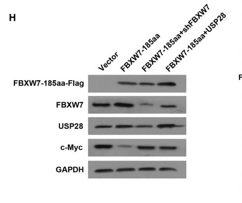
    Application: WB Species: human Sample: HEK293A cells

    Fig. 6.| SIRT5 promotes ccRCC cell proliferation by interacting with and desuccinylating SDHA. (A-B) ACHN cells were transfected with scramble shRNA or SIRT5 shRNA plasmid, SDHA was precipitated and its succinylation level was probed by anti-pan-succinylation antibody. SIRT5 silencing led to hypersuccinylation of SDHA in ccRCC cells and increased activity of SDHA. (C-E) SIRT5 silencing decreased cellular NADPH/NADP+and GSH/GSSG ratio and increased cellular ROS level. (F) Co-IP of SIRT5 and FLAG-tagged WT SDHA in HEK293A cells revealed SIRT5 coimmunoprecipitated with SDHA. (G-J) SIRT5 silencing repressed ACHN and 769-P cell proliferation and colony formation. Methods followed Fig. 5G-J.

    6). Li Y et al. Neuropilin-2 Signaling Modulates Mossy Fiber Sprouting by Regulating Axon Collateral Formation Through CRMP2 in a Rat Model of Epilepsy. Mol Neurobiol 2022 Aug 31. (PubMed: 36044155) [IF=5.682]

    7). Sun L et al. Overexpression of lncRNA-Gm2044 in spermatogonia impairs spermatogenesis in partial seminiferous tubules. Int J Mol Med 2022 Feb;49(2):17. (PubMed: 34935058) [IF=5.314]

    8). Wang C et al. Gankyrin activates the hedgehog signalling to drive metastasis in osteosarcoma. J Cell Mol Med 2021 Jun 5. (PubMed: 34089292) [IF=5.295]
    产品细节图片5

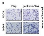
    Application: IHC Species: Human Sample: U2OS and MG63 cells

    FIGURE 1 Gankyrin promotes migration and invasion in OS in vitro. A, Induction of gankyrin in U2OS and MG63 cells was confirmed by Western blot analysis. B, Knockdown of gankyrin in U2OS and MG63 cells was confirmed via Western blot analysis. C, Induction of gankyrin significantly increased OS cell migration ability, as shown by wound healing assays. *P < .05; **P < .01. D, Up‐regulated gankyrin remarkably promoted OS cell invasion, as shown by Transwell invasion assays. *P < .05. E, Knockdown of gankyrin reduced OS cell migration ability. **P < .01. F, Knockdown of gankyrin inhibited OS cell invasion. **P < .01; ***P < .001

    9). Zhang Y et al. Treatment of diabetes mellitus-induced erectile dysfunction using endothelial progenitor cells genetically modified with human telomerase reverse transcriptase. Oncotarget 2016 Jun 28;7(26):39302-39315 (PubMed: 27283992)

    10). Xu F et al. Ablation of Cbl-b and c-Cbl in dendritic cells causes spontaneous liver cirrhosis via altering multiple properties of CD103+ cDC1s. Cell Death Discov 2022 Mar 30;8(1):142. (PubMed: 35354799)
    产品细节图片6

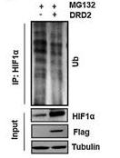
    Application: WB Species: human Sample: HEK293T cells

    Fig. 6 |Cbl-b and c-Cbl ubiquitinate different lysine sites of STAT5. The ubiquitination of STAT5a (A) and STAT5b (B) by Cbl-b and c-Cbl in HEK293T cells.

    风险提示:丁香通仅作为第三方平台,为商家信息发布提供平台空间。用户咨询产品时请注意保护个人信息及财产安全,合理判断,谨慎选购商品,商家和用户对交易行为负责。对于医疗器械类产品,请先查证核实企业经营资质和医疗器械产品注册证情况。

    图标文献和实验
    相关实验
    • 对比 Strep-tag 与 His-tag 系统异同点?

      质,小分子或是金属。当融合有亲和标签的蛋白通过标签-配体作用结合再层析基质上时,通过洗杂步骤,即可去除掉其他的细胞成分。 为了洗脱所需要的蛋白质,通过改变缓冲液的条件,如pH,或通过竞争亲和标签和配体连接的方法来进行。 但怎样的纯化是较为成功的呢?仅达到所需的质量量级,如毫克级(或克级)即可满足吗? 这些并不简单。质在蛋白纯化中同样尤为重要,比如,就蛋白的生物活性和纯度而言,通常会有所要求。   以下我们来讨论并且比较2种技术: His-tag系统和Strep-tag®系统,都使用了亲和层析法来纯化

    • Strep-tag 0 基础如何入门?

      Strep-tag®技术是亲和纯化重组蛋白的常用工具,它基于自然界中最强的非共价相互作用之一,即生物素与链霉亲和素的相互作用。该纯化系统的突出特点是纯化过程温和、目的蛋白纯度高、特异性强、兼容多种表达系统,对于有挑战性蛋白的纯化十分有帮助,因此近些年来脱颖而出。   本文将从以下方面介绍Strep-tag®技术: 标签介绍 纯化原理 标签-配体 纯化步骤 系统特点 标签介绍 目前有2种广泛使用的Strep标签: 它们分别是: Strep-tag®II 8 aa小标签(Trp-Ser

    • 在免疫沉淀IP免疫荧光IF如何正确的选择Flag标签抗体

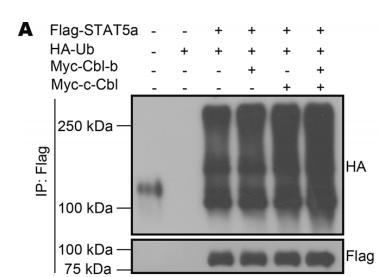
      融合表达蛋白等。比如在免疫荧光IF的实验里,同时配合EarthOx的Dylight 549荧光二抗,使用该抗体可以准确的定位出Flag融合表达蛋白在细胞内的位置(如图红色显示部分)。另外,做融合表达分析前,通过免疫共沉淀技术获得Flag融合表达蛋白也是非常重要的一步,因此选择一个合适的、可用于免疫共沉淀(Immunoprecipitation,IP)Flag标签抗体也是很必要的。EarthOx的Anti-Flag Tag Monoclonal Antibody(货号#E022060)不仅能应用

    图标技术资料

    暂无技术资料 索取技术资料

    同类产品报价

    产品名称
    产品价格
    公司名称
    报价日期
    ¥1
    上海信裕生物科技有限公司
    2025年07月11日询价
    询价
    上海沪震实业有限公司
    2025年07月09日询价
    ¥899
    南京安研生物科技有限公司
    2025年12月10日询价
    ¥1380
    上海允麦生物科技有限公司
    2025年12月12日询价
    询价
    上海远慕生物科技有限公司
    2025年07月13日询价
    Flag Tag标签抗体
    ¥1250