


- ¥1380 - 2200
- LMAI Bio
- LM-7362R
- 进口/国产
- 2026年03月10日
- WB=1:500-2000 ELISA=1:500-1000 IHC-P=1:400-800 IHC-F=1:400-800 ICC=1:100-500 IF=1:100-500 (石蜡切片需做抗原修复)
- Rabbit
- Human, Mouse, Rat,

企业认证
相关产品推荐更多 >


万千商家帮你免费找货
0 人在求购买到急需产品
- 详细信息
- 文献和实验
- 技术资料
- 供应商:
上海联迈生物工程有限公司
- 库存:
大量
- 目录编号:
LM-7362R
- 克隆性:
多克隆
- 抗原来源:
Rabbit
- 保质期:
1年
- 抗体英文名:
Melan A
- 抗体名:
黑色素瘤相关抗原/黑色素-A抗体
- 宿主:
Rabbit
- 适应物种:
Human, Mouse, Rat,
- 免疫原:
KLH conjugated synthetic peptide derived from mouse Melan A:1-80/113
- 亚型:
IgG
- 形态:
Lyophilized or Liquid
- 应用范围:
WB=1:500-2000 ELISA=1:500-1000 IHC-P=1:400-800 IHC-F=1:400-800 ICC=1:100-500 IF=1:100-500 (石蜡切片需做抗原修复)
- 浓度:
1mg/ml
- 保存条件:
Store at -20 °C
- 规格:
100ul 200ul
| 英文名称 | Melan A |
| 中文名称 | 黑色素瘤相关抗原/黑色素-A抗体 |
| 别 名 | Protein Melan-A; Antigen LB39 AA; Melanoma HMB45; Antigen SK29 AA; Antigen SK29-AA; CMM 1; CMM; CMM1; Cutaneous Malignant Melanoma Dysplastic Nevus; DNS; Dysplastic Nevus Syndrome; FAMMM; MART1; melan A; Melan A protein; Melanoma antigen recognized by T-cells 1; MLM; Monophenol monooxygenase; Tumor rejection antigen AB; tyrosinase; Melanoma HMB45; Melanoma; Melan-A; MART-1; MAR1_HUMAN. |
| 规格价格 | 100ul/1380元 购买 200ul/2200元 购买 大包装/询价 |
| 说 明 书 | 100ul 200ul |
| 研究领域 | 肿瘤 细胞生物 免疫学 t-淋巴细胞 |
| 抗体来源 | Rabbit |
| 克隆类型 | Polyclonal |
| 交叉反应 | Human, Mouse, Rat, |
| 产品应用 | WB=1:500-2000 ELISA=1:500-1000 IHC-P=1:400-800 IHC-F=1:400-800 ICC=1:100-500 IF=1:100-500 (石蜡切片需做抗原修复) not yet tested in other applications. optimal dilutions/concentrations should be determined by the end user. |
| 分 子 量 | 13kDa |
| 细胞定位 | 细胞浆 |
| 性 状 | Lyophilized or Liquid |
| 浓 度 | 1mg/ml |
| 免 疫 原 | KLH conjugated synthetic peptide derived from mouse Melan A:1-80/113 |
| 亚 型 | IgG |
| 纯化方法 | affinity purified by Protein A |
| 储 存 液 | 0.01M TBS(pH7.4) with 1% BSA, 0.03% Proclin300 and 50% Glycerol. |
| 保存条件 | Store at -20 °C for one year. Avoid repeated freeze/thaw cycles. The lyophilized antibody is stable at room temperature for at least one month and for greater than a year when kept at -20°C. When reconstituted in sterile pH 7.4 0.01M PBS or diluent of antibody the antibody is stable for at least two weeks at 2-4 °C. |
| PubMed | PubMed |
| 产品介绍 | background: Melanoma-associated antigens recognized by cytotoxic T lymphocytes (CTL) have been grouped into three categories: melanocyte differentiation antigens, cancer/testis-specific antigens and mutated or aberrantly expressed antigens. Many of these antigens consist of peptides that are presented to T cells by HLA molecules; they represent potential targets for cancer immunotherapy. Melan-A (also designated MART-1) is a melanocyte differentiation antigen that is specific to melanomas, melanocyte cell lines and retina. Melan-A peptide is recognized by most HLA-A2-restricted tumor-specific tumor-infiltrating lymphocytes in patients with melanoma. Antimelanoma cytotoxic T lymphocytes can be generated with a Melan-A peptide, implicating Melan-A as a potential candidate for antigen-specific immunotherapy in melanoma patients. Function: Involved in melanosome biogenesis by ensuring the stability of GPR143. Plays a vital role in the expression, stability, trafficking, and processing of melanocyte protein PMEL, which is critical to the formation of stage II melanosomes. Subunit: Interacts with PMEL. Interacts with GPR143. Subcellular Location: Endoplasmic reticulum membrane. Golgi apparatus. Golgi apparatus > trans-Golgi network membrane. Melanosome. Also found in small vesicles and tubules dispersed over the entire cytoplasm. A small fraction of the protein is inserted into the membrane in an inverted orientation. Inversion of membrane topology results in the relocalization of the protein from a predominant Endoplasmic reticulum membrane. Golgi apparatus. Golgi apparatus > trans-Golgi network membrane. Melanosome. Also found in small vesicles and tubules dispersed over the entire cytoplasm. A small fraction of the protein is inserted into the membrane in an inverted orientation. Inversion of membrane topology results in the relocalization of the protein from a predominant Golgi/post-Golgi area to the endoplasmic reticulum. Melanoma cells expressing the protein with an inverted membrane topology are more effectively recognized by specific cytolytic T-lymphocytes than those expressing the protein in its native membrane orientation. Tissue Specificity: Expression is restricted to melanoma and melanocyte cell lines and retina. Post-translational modifications: Acylated. SWISS: Q2TA50 Gene ID: 77836 Database links: Entrez Gene: 77836 Mouse Entrez Gene: 293890 Rat Important Note: This product as supplied is intended for research use only, not for use in human, therapeutic or diagnostic applications. |
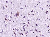
| 产品图片 | 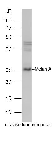 Protein: disease lung in mouse lysate; Primary: rabbit Anti-Melan A (bs-7362R) at 1:300; Secondary: HRP conjugated Goat-Anti-rabbit IgG(bs-0295G-HRP) at 1: 5000; Predicted band size: 26 kD Observed band size: 26 kD 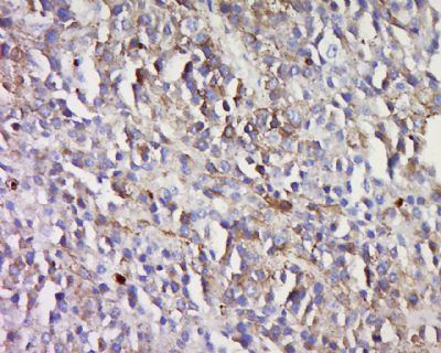 Tissue/cell: human melanoma tissue; 4% Paraformaldehyde-fixed and paraffin-embedded; Antigen retrieval: citrate buffer ( 0.01M, pH 6.0 ), Boiling bathing for 15min; Block endogenous peroxidase by 3% Hydrogen peroxide for 30min; Blocking buffer (normal goat serum,C-0005) at 37℃ for 20 min; Incubation: Anti-Melan A Polyclonal Antibody, Unconjugated(bs-7362R) 1:500, overnight at 4°C, followed by conjugation to the secondary antibody(SP-0023) and DAB(C-0010) staining |
风险提示:丁香通仅作为第三方平台,为商家信息发布提供平台空间。用户咨询产品时请注意保护个人信息及财产安全,合理判断,谨慎选购商品,商家和用户对交易行为负责。对于医疗器械类产品,请先查证核实企业经营资质和医疗器械产品注册证情况。
 文献和实验
文献和实验Nature 重磅:肿瘤微生物来源的多肽可作为免疫治疗的新靶点
等人 Nature 在线发表题为 Identification of bacteria-derived HLA-bound peptides in melanoma 研究论文。研究发现入侵肿瘤细胞的细菌会将其蛋白片段呈递到肿瘤细胞表面,随后被免疫系统识别,这一发现将可能被用于癌症的免疫疗法。 图片来源:Nature黑色素瘤是一种皮肤癌症,有三种已知的肿瘤相关抗原,它的细胞通常携带许多基因突变,很容易导致新的肿瘤抗原出现。因此研究团队以黑色素瘤样本来进行肿瘤抗原的探究。研究人员调查了 9 个人中 17 个黑色素
癌症疫苗全新设计——球形核酸疫苗!已在 7 种癌症中研究,曾应用于新冠,效果惊艳
%,并在动物模型中减缓了肿瘤的生长。 该方法为重新开发癌症疫苗和其他疫苗(包括 COVID-19 等传染病疫苗)绘制了新的蓝图。 图 1:来源 Nature Biomedical Engineering 研究方法与内容 在攻克癌症的众多疗法中,通过接种疫苗对抗表达靶向肿瘤相关抗原和新抗原的癌细胞,是一种独具吸引力的抗癌策略。以黑素瘤为例,科学家们致力于开发靶向肿瘤相关蛋白 (如 gp100、MAGE-A3、MART-1 和 NY-ESO-1) 的癌症疫苗。这些疫苗可以增加激活的黑色素瘤特异
来源、不同分化阶段的细胞可表达不同的分化抗原。利用人黑色素瘤特异的CTL克隆已鉴定出多种黑色素细胞分化抗原,这些抗原在多种黑色素瘤细胞呈异常表达,但在正常黑色素细胞仅呈轻微表达。现已发现的Pmel17 gpl00、酪氨酸酶和Melan-AMART-1 等分化抗原在黑色素瘤细胞中呈高表达,而且不同患者的黑色素瘤的分化抗原的结构高度同源,即很少显示个体差异,其机制可能涉及黑色素瘤细胞在生长发育的特定阶段,发生基因的异常性激活或调节基因发生突变,引起编码蛋白异常表达和细胞恶性转化。这些异常表达的分化
 技术资料
技术资料暂无技术资料 索取技术资料

