

- ¥1380 - 2200
- LMAI Bio
- LM-14045R
- 进口/国产
- 2026年06月15日
- ELISA=1:500-1000 IHC-P=1:400-800 IHC-F=1:400-800 ICC=1:100-500 IF=1:100-500 (石蜡切片需做抗原修复)
- Rabbit
- Human

企业认证
相关产品推荐更多 >
万千商家帮你免费找货
0 人在求购买到急需产品
- 详细信息
- 文献和实验
- 技术资料
- 供应商:
上海联迈生物工程有限公司
- 库存:
大量
- 目录编号:
LM-14045R
- 克隆性:
多克隆
- 抗原来源:
Rabbit
- 保质期:
1年
- 抗体英文名:
CRB1
- 抗体名:
CRB1蛋白抗体
- 宿主:
Rabbit
- 适应物种:
Human
- 免疫原:
KLH conjugated synthetic peptide derived from human CRB1:301-400/1406 <Extracellular>
- 亚型:
IgG
- 形态:
Lyophilized or Liquid
- 应用范围:
ELISA=1:500-1000 IHC-P=1:400-800 IHC-F=1:400-800 ICC=1:100-500 IF=1:100-500 (石蜡切片需做抗原修复)
- 浓度:
1mg/ml
- 保存条件:
Store at -20 °C
- 规格:
100ul 200ul
| 英文名称 | CRB1 |
| 中文名称 | CRB1蛋白抗体 |
| 别 名 | CRB1; CRUM1_HUMAN; Protein crumbs homolog 1. |
| 规格价格 | 100ul/1380元 购买 200ul/2200元 购买 大包装/询价 |
| 说 明 书 | 100ul 200ul |
| 研究领域 | 细胞生物 神经生物学 细胞骨架 |
| 抗体来源 | Rabbit |
| 克隆类型 | Polyclonal |
| 交叉反应 | Human, |
| 产品应用 | ELISA=1:500-1000 IHC-P=1:400-800 IHC-F=1:400-800 ICC=1:100-500 IF=1:100-500 (石蜡切片需做抗原修复) not yet tested in other applications. optimal dilutions/concentrations should be determined by the end user. |
| 分 子 量 | 151kDa |
| 细胞定位 | 细胞浆 细胞膜 |
| 性 状 | Lyophilized or Liquid |
| 浓 度 | 1mg/ml |
| 免 疫 原 | KLH conjugated synthetic peptide derived from human CRB1:301-400/1406 <Extracellular> |
| 亚 型 | IgG |
| 纯化方法 | affinity purified by Protein A |
| 储 存 液 | 0.01M TBS(pH7.4) with 1% BSA, 0.03% Proclin300 and 50% Glycerol. |
| 保存条件 | Store at -20 °C for one year. Avoid repeated freeze/thaw cycles. The lyophilized antibody is stable at room temperature for at least one month and for greater than a year when kept at -20°C. When reconstituted in sterile pH 7.4 0.01M PBS or diluent of antibody the antibody is stable for at least two weeks at 2-4 °C. |
| PubMed | PubMed |
| 产品介绍 | background: This gene encodes a protein which is similar to the Drosophila crumbs protein and localizes to the inner segment of mammalian photoreceptors. In Drosophila crumbs localizes to the stalk of the fly photoreceptor and may be a component of the molecular scaffold that controls proper development of polarity in the eye. Mutations in this gene are associated with a severe form of retinitis pigmentosa, RP12, and with Leber congenital amaurosis. Alternate splicing results in multiple transcript variants, some protein coding and some non-protein coding.[provided by RefSeq, Apr 2012] Function: Plays a role in photoreceptor morphogenesis in the retina. May maintain cell polarization and adhesion. Subcellular Location: Secreted and Apical cell membrane. Distributed at the apical membrane of all retinal epithelial cells. Located in the apical membrane of the adherens junction in outer limiting membrane (OLM) of the retina. Tissue Specificity: Preferential expression in retina, also expressed in brain, testis, fetal brain and fetal eye. Post-translational modifications: Extensively glycosylated. DISEASE: Note=CRB1 mutations have been found in various retinal dystrophies, chronic and disabling disorders of visual function. They predominantly involve the posterior portion of the ocular fundus, due to degeneration in the sensory layer of the retina, retinal pigment epithelium, Bruch membrane, choroid, or a combination of these tissues. Onset of inherited retinal dystrophies is painless, bilateral and typically progressive. Most people experience gradual peripheral vision loss or tunnel vision, and difficulties with poor illumination and night vision. Central vision is usually unaffected, so the person may still be able to read. However, it can also deteriorate to cause total blindness. Examples of retinal dystrophies are retinitis pigmentosa, Leber congenital amaurosis, cone-rod dystrophy among others. Defects in CRB1 are the cause of retinitis pigmentosa type 12 (RP12) [MIM:600105]. A retinal dystrophy belonging to the group of pigmentary retinopathies. Retinitis pigmentosa is characterized by retinal pigment deposits visible on fundus examination and primary loss of rod photoreceptor cells, followed by secondary loss of cone photoreceptors. Patients typically have night vision blindness and loss of midperipheral visual field. As their condition progresses, they lose their far peripheral visual field and eventually central vision as well. RP12 is an autosomal recessive severe form oFTen manifesting in early childhood. Patients experiment progressive visual field loss with severe visual impairment before the age of twenty. Some patients have a preserved paraarteriolar retinal pigment epithelium (PPRPE) and hypermetropia. Defects in CRB1 are the cause of Leber congenital amaurosis type 8 (LCA8) [MIM:613835]. LCA designates a clinically and genetically heterogeneous group of childhood retinal degenerations, generally inherited in an autosomal recessive manner. Affected infants have little or no retinal photoreceptor function as tested by electroretinography. LCA represents the most common genetic cause of congenital visual impairment in infants and children. Defects in CRB1 are the cause of pigmented paravenous chorioretinal atrophy (PPCRA) [MIM:172870]. PPCRA is an unusual retinal degeneration characterized by accumulation of pigmentation along retinal veins. PPCRA is dominantly inherited, but exhibited variable expressivity. Males are more likely to exhibit a severe phenotype, whereas females may remain virtually asymptomatic even in later years. The PPCRA phenotype is associated with a mutation in CRB1 gene which is likely to affect the structure of the CRB1 protein. Similarity: Belongs to the Crumbs protein family. Contains 19 EGF-like domains. Contains 3 laminin G-like domains. SWISS: P82279 Gene ID: 23418 Database links: Entrez Gene: 23418 Human SwissProt: P82279 Human Unigene: 126135 Human Important Note: This product as supplied is intended for research use only, not for use in human, therapeutic or diagnostic applications. |
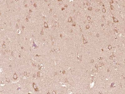
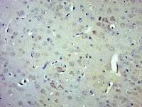
| 产品图片 |  Paraformaldehyde-fixed, paraffin embedded (Human brain glioma); Antigen retrieval by boiling in sodium citrate buffer (pH6.0) for 15min; Block endogenous peroxidase by 3% hydrogen peroxide for 20 minutes; Blocking buffer (normal goat serum) at 37°C for 30min; Antibody incubation with (CRB1) Polyclonal Antibody, Unconjugated (bs-14045R) at 1:400 overnight at 4°C, followed by operating according to SP Kit(Rabbit) (sp-0023) instructionsand DAB staining. |
风险提示:丁香通仅作为第三方平台,为商家信息发布提供平台空间。用户咨询产品时请注意保护个人信息及财产安全,合理判断,谨慎选购商品,商家和用户对交易行为负责。对于医疗器械类产品,请先查证核实企业经营资质和医疗器械产品注册证情况。
 文献和实验
文献和实验人 抗麦胶蛋白抗体 ( AGA) 酶联免疫分析 试剂盒使用说明书 本试剂仅供研究使用 目的:本试剂盒用于测定人血清,血浆及相关液体样本中 抗麦胶蛋白抗体( AGA) 的 含量。 实验原理: 本试剂盒应用双抗原夹心法测定标本中人 抗麦胶蛋白抗体( AGA) 的 水平。用纯化的 抗原 包被微孔板,制成固相抗原,往包被抗原的微孔中依次加入 抗麦胶蛋白抗体( AGA) ,再与 HRP 标记的 抗原 结合
sunchangzheng 我打算用此抗体通过流式检测细胞表面P糖蛋白的表达量差异。 说明书中说是多抗,这种抗体有单抗的吧? 用于FCM: 1:20-100,这是说用于流式的稀释倍数,是不是稀释倍数越小越好呢? 同型对照抗体是不是必须要做的?如何选择阴型同型对照抗体呢! 我用的抗体是FITC标记的P-糖蛋白抗体 谢谢交流与帮助! freecell 抗体的稀释倍数是需要优化
载脂蛋白抗体的制备 抗原免疫动物产生的抗体是血清γ-球蛋白的一部分。抗体的理化性质及结构与γ-球蛋白相近似。γ-球蛋白是一组结构相似,但又有差异的蛋白质,通称为免疫球蛋白(immunoglobulin,Ig),按其结构和免疫化学性质的差异,又可分为五类,分别称为IgG、IgA、IgM、IgD和IgE。载脂蛋白抗体以IgG最为稳定。因载脂蛋白的体液中含量不同以及分离纯化的难易的差别,可分别采用多克隆抗体制备法和单克隆抗体制备法。 1.多克隆抗体的制备 (1)抗原
 技术资料
技术资料暂无技术资料 索取技术资料

