


- ¥780 - 2200
- LMAI Bio
- LM-0908R
- 进口/国产
- 2026年05月27日
- ELISA=1:500-1000 IHC-P=1:400-800 IHC-F=1:400-800 Flow-Cyt=1μg/Test IF=1:100-500 (石蜡切片需做抗原修复)
- Rabbit
- Human, Mouse, Rat, Chicken, Dog, Pig, Cow,

企业认证
相关产品推荐更多 >
万千商家帮你免费找货
0 人在求购买到急需产品
- 详细信息
- 文献和实验
- 技术资料
- 供应商:
上海联迈生物工程有限公司
- 库存:
大量
- 目录编号:
LM-0908R
- 克隆性:
多克隆
- 抗原来源:
Rabbit
- 保质期:
1年
- 抗体英文名:
JAK2
- 抗体名:
蛋白质酪氨酸激酶JAK2抗体
- 宿主:
Rabbit
- 适应物种:
Human, Mouse, Rat, Chicken, Dog, Pig, Cow,
- 免疫原:
KLH conjugated synthetic peptide derived from human JAK2:601-700/1132
- 亚型:
IgG
- 形态:
Lyophilized or Liquid
- 应用范围:
ELISA=1:500-1000 IHC-P=1:400-800 IHC-F=1:400-800 Flow-Cyt=1μg/Test IF=1:100-500 (石蜡切片需做抗原修复)
- 浓度:
1mg/ml
- 保存条件:
Store at -20 °C
- 规格:
50ul 100ul 200ul
| 英文名称 | JAK2 |
| 中文名称 | 蛋白质酪氨酸激酶JAK2抗体 |
| 别 名 | Tyrosine protein kinase JAK2; JAK 2; JAK-2; JAK2; JAK2_HUMAN; Janus Activating Kinase 2; Janus Kinase 2; JTK 10; JTK10; OTTHUMP00000043260; Tyrosine-protein kinase JAK2; Tyrosine protein kinase JAK2. |
 |
Specific References (1) | bs-0908R has been referenced in 1 publications. [IF=0.82] Wei, Yanfei, et al. "Plumbagin Inhibits Leptin-Induced Proliferation of Hepatic Stellate Cells via JAK2-STAT3 Pathway to Protect against Hepatic Fibrosis."Tropical Journal of Pharmaceutical Research?12.5 (2013): 691-698. WB ; Human. PubMed:N/A |
| 规格价格 | 50ul/780元 购买 100ul/1380元 购买 200ul/2200元 购买 大包装/询价 |
| 说 明 书 | 50ul 100ul 200ul |
| 研究领域 | 肿瘤 细胞生物 免疫学 染色质和核信号 信号转导 激酶和磷酸酶 表观遗传学 |
| 抗体来源 | Rabbit |
| 克隆类型 | Polyclonal |
| 交叉反应 | Human, Mouse, Rat, Chicken, Dog, Pig, Cow, |
| 产品应用 | ELISA=1:500-1000 IHC-P=1:400-800 IHC-F=1:400-800 Flow-Cyt=1μg/Test IF=1:100-500 (石蜡切片需做抗原修复) not yet tested in other applications. optimal dilutions/concentrations should be determined by the end user. |
| 分 子 量 | 131kDa |
| 细胞定位 | 细胞核 细胞浆 细胞膜 |
| 性 状 | Lyophilized or Liquid |
| 浓 度 | 1mg/ml |
| 免 疫 原 | KLH conjugated synthetic peptide derived from human JAK2:601-700/1132 |
| 亚 型 | IgG |
| 纯化方法 | affinity purified by Protein A |
| 储 存 液 | 0.01M TBS(pH7.4) with 1% BSA, 0.03% Proclin300 and 50% Glycerol. |
| 保存条件 | Store at -20 °C for one year. Avoid repeated freeze/thaw cycles. The lyophilized antibody is stable at room temperature for at least one month and for greater than a year when kept at -20°C. When reconstituted in sterile pH 7.4 0.01M PBS or diluent of antibody the antibody is stable for at least two weeks at 2-4 °C. |
| PubMed | PubMed |
| 产品介绍 | background: This gene product is a protein tyrosine kinase involved in a specific subset of cytokine receptor signaling pathways. It has been found to be constituitively associated with the prolactin receptor and is required for responses to gamma interferon. Mice that do not express an active protein for this gene exhibit embryonic lethality associated with the absence of definitive erythropoiesis. [provided by RefSeq, Jul 2008] Function: Non-receptor tyrosine kinase involved in various processes such as cell growth, development, differentiation or histone modifications. Mediates essential signaling events in both innate and adaptive immunity. In the cytoplasm, plays a pivotal role in signal transduction via its association with type I receptors such as growth hormone (GHR), prolactin (PRLR), leptin (LEPR), erythropoietin (EPOR), thrombopoietin (THPO); or type II receptors including IFN-alpha, IFN-beta, IFN-gamma and multiple interleukins. Following ligand-binding to cell surface receptors, phosphorylates specific tyrosine residues on the cytoplasmic tails of the receptor, creating docking sites for STATs proteins. Subsequently, phosphorylates the STATs proteins once they are recruited to the receptor. Phosphorylated STATs then form homodimer or heterodimers and translocate to the nucleus to activate gene transcription. For example, cell stimulation with erythropoietin (EPO) during erythropoiesis leads to JAK2 autophosphorylation, activation, and its association with erythropoietin receptor (EPOR) that becomes phosphorylated in its cytoplasmic domain. Then, STAT5 (STAT5A or STAT5B) is recruited, phosphorylated and activated by JAK2. Once activated, dimerized STAT5 translocates into the nucleus and promotes the transcription of several essential genes involved in the modulation of erythropoiesis. In addition, JAK2 mediates angiotensin-2-induced ARHGEF1 phosphorylation. Plays a role in cell cycle by phosphorylating CDKN1B. Cooperates with TEC through reciprocal phosphorylation to mediate cytokine-driven activation of FOS transcription. In the nucleus, plays a key role in chromatin by specifically mediating phosphorylation of 'Tyr-41' of histone H3 (H3Y41ph), a specific tag that promotes exclusion of CBX5 (HP1 alpha) from chromatin. Subunit: Interacts with EPOR, LYN, SIRPA, SH2B1 and TEC (By similarity). Interacts with IL23R, SKB1 and STAM2. Subcellular Location: Endomembrane system; Peripheral membrane protein (By similarity). Cytoplasm. Nucleus. Tissue Specificity: Ubiquitously expressed throughout most tissues. Post-translational modifications: Autophosphorylated, leading to regulate its activity. Leptin promotes phosphorylation on tyrosine residues, including phosphorylation on Tyr-813. Autophosphorylation on Tyr-119 in response to EPO down-regulates its kinase activity. Autophosphorylation on Tyr-868, Tyr-966 and Tyr-972 in response to growth hormone (GH) are required for maximal kinase activity. Also phosphorylated by TEC. DISEASE: Note=Chromosomal aberrations involving JAK2 are found in both chronic and acute forms of eosinophilic, lymphoblastic and myeloid leukemia. Translocation t(8;9)(p22;p24) with PCM1 links the protein kinase domain of JAK2 to the major portion of PCM1. Translocation t(9;12)(p24;p13) with ETV6. Defects in JAK2 are a cause of susceptibility to Budd-Chiari syndrome (BDCHS) [MIM:600880]. A syndrome caused by obstruction of hepatic venous outflow involving either the hepatic veins or the terminal segment of the inferior vena cava. Obstructions are generally caused by thrombosis and lead to hepatic congestion and ischemic necrosis. Clinical manifestations observed in the majority of patients include hepatomegaly, right upper quadrant pain and abdominal ascites. Budd-Chiari syndrome is associated with a combination of disease states including primary myeloproliferative syndromes and thrombophilia due to factor V Leiden, protein C deficiency and antithrombin III deficiency. Budd-Chiari syndrome is a rare but typical complication in patients with polycythemia vera. Similarity: Belongs to the protein kinase superfamily. Tyr protein kinase family. JAK subfamily. Contains 1 FERM domain. Contains 1 protein kinase domain. Contains 1 SH2 domain. SWISS: O60674 Gene ID: 3717 Database links: Entrez Gene: 3717 Human Entrez Gene: 16452 Mouse Entrez Gene: 24514 Rat GenBank: NP_004963 Human Omim: 147796 Human SwissProt: O60674 Human SwissProt: Q62120 Mouse SwissProt: Q62689 Rat Unigene: 656213 Human Unigene: 275839 Mouse Unigene: 18909 Rat Important Note: This product as supplied is intended for research use only, not for use in human, therapeutic or diagnostic applications. |
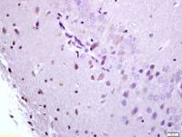
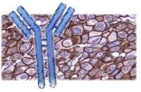
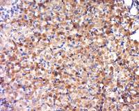
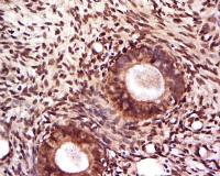
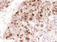
| 产品图片 | 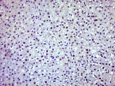 Paraformaldehyde-fixed, paraffin embedded (Rat liver); Antigen retrieval by boiling in sodium citrate buffer (pH6.0) for 15min; Block endogenous peroxidase by 3% hydrogen peroxide for 20 minutes; Blocking buffer (normal goat serum) at 37°C for 30min; Antibody incubation with (JAK2) Polyclonal Antibody, Unconjugated (bs-0908R) at 1:500 overnight at 4°C, followed by a conjugated secondary (sp-0023) for 20 minutes and DAB staining. 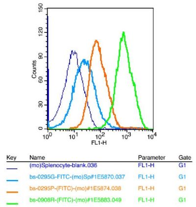 Blank control: mouse splenocytes(blue) Isotype Control Antibody: Rabbit IgG(orange) ; Secondary Antibody: Goat anti-rabbit IgG-FITC(white blue), Dilution: 1:100 in 1 X PBS containing 0.5% BSA ; Primary Antibody Dilution: 1μl in 100 μL1X PBS containing 0.5% BSA(green). |
风险提示:丁香通仅作为第三方平台,为商家信息发布提供平台空间。用户咨询产品时请注意保护个人信息及财产安全,合理判断,谨慎选购商品,商家和用户对交易行为负责。对于医疗器械类产品,请先查证核实企业经营资质和医疗器械产品注册证情况。
 文献和实验
文献和实验受体相关性蛋白酪氨酸激酶介导的信号途径 大部分细胞因子受体分子胞内区不带有启动信号转导的蛋白酪氨酸激酶(PTK)结构域,但可通过相应胞内区连接的PTK起作用。CkR―F和IFNR―F的受体分子胞内区都结合有PTK中的Janus激酶(Janus kinase,Jak)家族成员,包括Jakl、Jak2、Jak3和Tyk2。细胞因子各种受体的亚单位往往连接Jak家族中的一个或几个特定成员。如C―CSFR与Jak2连接,IL2Rβ链与Jakl、IL2Rγ链与Jak3连接,ILl0R
尼古丁乙酰胆碱受体(nAChRs)出现在神经细胞和免疫细胞中。研究人员在8月出版《自然-免疫学》上说,nAChRs能启动一个可关闭免疫细胞发炎机制的程序。这种触发器可用于防止发炎。新工作进一步提供证据表明:神经系统与免疫系统有密切关系。巨噬细胞是免疫细胞,它能产生大量的蛋白质和化学物质从而引起发炎效应。Wouter de Jonge和同事发现,触发巨噬细胞上的nAChRs能启动细胞中名为Jak2和 STAT3的小分子,它们可减少防御蛋白质产量从而降低发炎的程度。为了展示这一发
的TF1细胞中检测出分子量97/95kDa的蛋白发生了酪氨酸磷酸化,抗gp130的信号转导中很重要。在不同的细胞系3T3-L1、B细胞杂交瘤、髓样 白血病 系中发现有不同分子量蛋白的要酪氨酸磷酸化,提示在不同的细胞系中存在细胞特异的酪氨酸激酶及各自特异的底物,这可能是共用gp130的IL-6、IL-11、LIF、CNTF、OSM在不同细胞中生物学作用差异的原因一。JAK2是一种非受体型的酪氨酸激酶,可以被EPO、IL-3、G-CSF、IL-6等多种细胞因子刺激所激活,JAK2可能
 技术资料
技术资料暂无技术资料 索取技术资料

