


- ¥780 - 2200
- LMAI Bio
- LM-0115R
- 进口/国产
- 2026年05月27日
- WB=1:500-2000 ELISA=1:500-1000 IHC-P=1:400-800 IHC-F=1:400-800 Flow-Cyt=1μg/Test IF=1:100-500 (石蜡切片需做抗原修复)
- Rabbit
- Human, Mouse, Rat, Chicken, Dog, Pig, Cow, Rabbit, Sheep,

企业认证
相关产品推荐更多 >

万千商家帮你免费找货
0 人在求购买到急需产品
- 详细信息
- 文献和实验
- 技术资料
- 供应商:
上海联迈生物工程有限公司
- 库存:
大量
- 目录编号:
LM-0115R
- 克隆性:
多克隆
- 抗原来源:
Rabbit
- 保质期:
1年
- 抗体英文名:
AKT1
- 抗体名:
蛋白激酶B抗体
- 宿主:
Rabbit
- 适应物种:
Human, Mouse, Rat, Chicken, Dog, Pig, Cow, Rabbit, Sheep,
- 免疫原:
KLH conjugated synthetic peptide derived from human PKB C-terminus:401-479/479
- 亚型:
IgG
- 形态:
Lyophilized or Liquid
- 应用范围:
WB=1:500-2000 ELISA=1:500-1000 IHC-P=1:400-800 IHC-F=1:400-800 Flow-Cyt=1μg/Test IF=1:100-500 (石蜡切片需做抗原修复)
- 浓度:
1mg/ml
- 保存条件:
Store at -20 °C
- 规格:
50ul 100ul 200ul
| 英文名称 | AKT1 |
| 中文名称 | 蛋白激酶B抗体 |
| 别 名 | AKT 1; AKT; AKT1; AKT-1; AKT1_HUMAN; C AKT; cAKT; MGC9965; MGC99656; Oncogene AKT1; PKB; PKB alpha; PKB-ALPHA; PRKBA; Protein Kinase B Alpha; Protein kinase B; Proto-oncogene c-Akt; RAC Alpha; RAC alpha serine/threonine protein kinase; RAC; RAC PK Alpha; Rac protein kinase alpha; RAC Serine/Threonine Protein Kinase; RAC-alpha serine/threonine-protein kinase; RAC-PK-alpha; v akt murine thymoma viral oncogene homolog 1; vAKT Murine Thymoma Viral Oncogene Homolog 1. |
 |
Specific References (4) | bs-0115R has been referenced in 4 publications. [IF=1.72] Li, Xinxin, et al. "Follistatin could promote the proliferation of duck primary myoblasts by activating PI3K/Akt/mTOR signaling." Bioscience Reports (2014). WB ; PubMed:25200144 [IF=2.56] Xu, C., et al. "Proteomics Analysis of Hepatocyte Proliferation Regulated by FGF, PDGF, Insulin, Oncostatin M and Interleukin 2 Signaling Pathways during Rat Liver Regeneration." J Proteomics Computational Biol 1.1 (2014): 8. WB ; Rat. PubMed:not posted yet [IF=5.23] Zhao, Yong, et al. "Hydrogen Sulfide and/or Ammonia Reduces Spermatozoa Motility through AMPK/AKT Related Pathways." Scientific Reports 6 (2016): 37884. WB ; Pig. PubMed:27883089 [IF=3.33] Xu, Hong-Wu, et al. "The expression of cytoglobin as a prognostic factor in gliomas: a retrospective analysis of 88 patients." BMC cancer 13.1 (2013): 247. IHC-P ; Human. PubMed:23688241 |
| 规格价格 | 50ul/780元 购买 100ul/1380元 购买 200ul/2200元 购买 大包装/询价 |
| 说 明 书 | 50ul 100ul 200ul |
| 研究领域 | 肿瘤 细胞生物 信号转导 细胞凋亡 激酶和磷酸酶 |
| 抗体来源 | Rabbit |
| 克隆类型 | Polyclonal |
| 交叉反应 | Human, Mouse, Rat, Chicken, Dog, Pig, Cow, Rabbit, Sheep, |
| 产品应用 | WB=1:500-2000 ELISA=1:500-1000 IHC-P=1:400-800 IHC-F=1:400-800 Flow-Cyt=1μg/Test IF=1:100-500 (石蜡切片需做抗原修复) not yet tested in other applications. optimal dilutions/concentrations should be determined by the end user. |
| 分 子 量 | 56kDa |
| 细胞定位 | 细胞核 细胞浆 细胞膜 |
| 性 状 | Lyophilized or Liquid |
| 浓 度 | 1mg/ml |
| 免 疫 原 | KLH conjugated synthetic peptide derived from human PKB C-terminus:401-479/479 |
| 亚 型 | IgG |
| 纯化方法 | affinity purified by Protein A |
| 储 存 液 | 0.01M TBS(pH7.4) with 1% BSA, 0.03% Proclin300 and 50% Glycerol. |
| 保存条件 | Store at -20 °C for one year. Avoid repeated freeze/thaw cycles. The lyophilized antibody is stable at room temperature for at least one month and for greater than a year when kept at -20°C. When reconstituted in sterile pH 7.4 0.01M PBS or diluent of antibody the antibody is stable for at least two weeks at 2-4 °C. |
| PubMed | PubMed |
| 产品介绍 | background: The serine-threonine protein kinase encoded by the AKT1 gene is catalytically inactive in serum-starved primary and immortalized fibroblasts. AKT1 and the related AKT2 are activated by platelet-derived growth factor. The activation is rapid and specific, and it is abrogated by mutations in the pleckstrin homology domain of AKT1. It was shown that the activation occurs through phosphatidylinositol 3-kinase. In the developing nervous system AKT is a critical mediator of growth factor-induced neuronal survival. Survival factors can suppress apoptosis in a transcription-independent manner by activating the serine/threonine kinase AKT1, which then phosphorylates and inactivates components of the apoptotic machinery. Mutations in this gene have been associated with the Proteus syndrome. Multiple alternatively spliced transcript variants have been found for this gene. [provided by RefSeq, Jul 2011] Function: AKT1 is one of 3 closely related serine/threonine-protein kinases (AKT1, AKT2 and AKT3) called the AKT kinase, and which regulate many processes including metabolism, proliferation, cell survival, growth and angiogenesis. This is mediated through serine and/or threonine phosphorylation of a range of downstream substrates. Over 100 substrate candidates have been reported so far, but for most of them, no isoform specificity has been reported. AKT is responsible of the regulation of glucose uptake by mediating insulin-induced translocation of the SLC2A4/GLUT4 glucose transporter to the cell surface. Phosphorylation of PTPN1 at 'Ser-50' negatively modulates its phosphatase activity preventing dephosphorylation of the insulin receptor and the attenuation of insulin signaling. Phosphorylation of TBC1D4 triggers the binding of this effector to inhibitory 14-3-3 proteins, which is required for insulin-stimulated glucose transport. AKT regulates also the storage of glucose in the form of glycogen by phosphorylating GSK3A at 'Ser-21' and GSK3B at 'Ser-9', resulting in inhibition of its kinase activity. Phosphorylation of GSK3 isoforms by AKT is also thought to be one mechanism by which cell proliferation is driven. AKT regulates also cell survival via the phosphorylation of MAP3K5 (apoptosis signal-related kinase). Phosphorylation of 'Ser-83' decreases MAP3K5 kinase activity stimulated by oxidative stress and thereby prevents apoptosis. AKT mediates insulin-stimulated protein synthesis by phosphorylating TSC2 at 'Ser-939' and 'Thr-1462', thereby activating mTORC1 signaling and leading to both phosphorylation of 4E-BP1 and in activation of RPS6KB1. AKT is involved in the phosphorylation of members of the FOXO factors (Forkhead family of transcription factors), leading to binding of 14-3-3 proteins and cytoplasmic localization. In particular, FOXO1 is phosphorylated at 'Thr-24', 'Ser-256' and 'Ser-319'. FOXO3 and FOXO4 are phosphorylated on equivalent sites. AKT has an important role in the regulation of NF-kappa-B-dependent gene transcription and positively regulates the activity of CREB1 (cyclic AMP (cAMP)-response element binding protein). The phosphorylation of CREB1 induces the binding of accessory proteins that are necessary for the transcription of pro-survival genes such as BCL2 and MCL1. AKT phosphorylates 'Ser-454' on ATP citrate lyase (ACLY), thereby potentially regulating ACLY activity and fatty acid synthesis. Activates the 3B isoform of cyclic nucleotide phosphodiesterase (PDE3B) via phosphorylation of 'Ser-273', resulting in reduced cyclic AMP levels and inhibition of lipolysis. Phosphorylates PIKFYVE on 'Ser-318', which results in increased PI(3)P-5 activity. The Rho GTPase-activating protein DLC1 is another substrate and its phosphorylation is implicated in the regulation cell proliferation and cell growth. AKT plays a role as key modulator of the AKT-mTOR signaling pathway controlling the tempo of the process of newborn neurons integration during adult neurogenesis, including correct neuron positioning, dendritic development and synapse formation. Signals downstream of phosphatidylinositol 3-kinase (PI(3)K) to mediate the effects of various growth factors such as platelet-derived growth factor (PDGF), epidermal growth factor (EGF), insulin and insulin-like growth factor I (IGF-I). AKT mediates the antiapoptotic effects of IGF-I. Essential for the SPATA13-mediated regulation of cell migration and adhesion assembly and disassembly. May be involved in the regulation of the placental development. Phosphorylates STK4/MST1 at 'Thr-120' and 'Thr-387' leading to inhibition of its: kinase activity, nuclear translocation, autophosphorylation and ability to phosphorylate FOXO3. Phosphorylates STK3/MST2 at 'Thr-117' and 'Thr-384' leading to inhibition of its: cleavage, kinase activity, autophosphorylation at Thr-180, binding to RASSF1 and nuclear translocation. Phosphorylates SRPK2 and enhances its kinase activity towards SRSF2 and ACIN1 and promotes its nuclear translocation. Phosphorylates RAF1 at 'Ser-259' and negatively regulates its activity. Phosphorylation of BAD stimulates its pro-apoptotic activity. Subunit: Interacts (via the C-terminus) with CCDC88A (via its C-terminus). Interacts with GRB10; the interaction leads to GRB10 phosphorylation thus promoting YWHAE-binding. Interacts with AGAP2 (isoform 2/PIKE-A); the interaction occurs in the presence of guanine nucleotides. Interacts with AKTIP. Interacts (via PH domain) with MTCP1, TCL1A AND TCL1B. Interacts with CDKN1B; the interaction phosphorylates CDKN1B promoting 14-3-3 binding and cell-cycle progression. Interacts with MAP3K5 and TRAF6. Interacts with BAD, PPP2R5B, STK3 and STK4. Interacts (via PH domain) with SIRT1. Interacts with SRPK2 in a phosphorylation-dependent manner. Interacts with RAF1. Interacts with TRIM13; the interaction ubiquitinates AKT1 leading to its proteasomal degradation. Interacts with TNK2 and CLK2. Interacts (via the C-terminus) with THEM4 (via its C-terminus). Interacts with and phosphorylated by PDPK1. Subcellular Location: Cytoplasm. Nucleus. Cell membrane. Note=Nucleus after activation by integrin-linked protein kinase 1 (ILK1). Nuclear translocation is enhanced by interaction with TCL1A. Phosphorylation on Tyr-176 by TNK2 results in its localization to the cell membrane where it is targeted for further phosphorylations on Thr-308 and Ser-473 leading to its activation and the activated form translocates to the nucleus. Tissue Specificity: Expressed in prostate cancer and levels increase from the normal to the malignant state (at protein level). Expressed in all human cell types so far analyzed. The Tyr-176 phosphorylated form shows a significant increase in expression in breast cancers during the progressive stages i.e. normal to hyperplasia (ADH), ductal carcinoma in situ (DCIS), invasive ductal carcinoma (IDC) and lymph node metastatic (LNMM) stages. Post-translational modifications: O-GlcNAcylation at Thr-305 and Thr-312 inhibits activating phosphorylation at Thr-308 via disrupting the interaction between AKT1 and PDPK1. O-GlcNAcylation at Ser-473 also probably interferes with phosphorylation at this site. Phosphorylation on Thr-308, Ser-473 and Tyr-474 is required for full activity. Activated TNK2 phosphorylates it on Tyr-176 resulting in its binding to the anionic plasma membrane phospholipid PA. This phosphorylated form localizes to the cell membrane, where it is targeted by PDPK1 and PDPK2 for further phosphorylations on Thr-308 and Ser-473 leading to its activation. Ser-473 phosphorylation by mTORC2 favors Thr-308 phosphorylation by PDPK1. Ser-473 phosphorylation is enhanced by interaction with AGAP2 isoform 2 (PIKE-A). Ser-473 phosphorylation is enhanced in focal cortical dysplasias with Taylor-type balloon cells. Ser-473 phosphorylation is enhanced by signaling through activated FLT3. Dephosphorylated at Thr-308 and Ser-473 by PP2A phosphatase. The phosphorylated form of PPP2R5B is required for bridging AKT1 with PP2A phosphatase. Ubiquitinated via 'Lys-48'-linked polyubiquitination by ZNRF1, leading to its degradation by the proteasome. Ubiquitinated; undergoes both 'Lys-48'- and 'Lys-63'-linked polyubiquitination. TRAF6-induced 'Lys-63'-linked AKT1 ubiquitination is critical for phosphorylation and activation. When ubiquitinated, it translocates to the plasma membrane, where it becomes phosphorylated. When fully phosphorylated and translocated into the nucleus, undergoes 'Lys-48'-polyubiquitination catalyzed by TTC3, leading to its degradation by the proteasome. Also ubiquitinated by TRIM13 leading to its proteasomal degradation. Acetylated on Lys-14 and Lys-20 by the histone acetyltransferases EP300 and KAT2B. Acetylation results in reduced phosphorylation and inhibition of activity. Deacetylated at Lys-14 and Lys-20 by SIRT1. SIRT1-mediated deacetylation relieves the inhibition. DISEASE: Defects in AKT1 are a cause of susceptibility to breast cancer (BC) [MIM:114480]. A common malignancy originating from breast epithelial tissue. Breast neoplasms can be distinguished by their histologic pattern. Invasive ductal carcinoma is by far the most common type. Breast cancer is etiologically and genetically heterogeneous. Important genetic factors have been indicated by familial occurrence and bilateral involvement. Mutations at more than one locus can be involved in different families or even in the same case. Defects in AKT1 are associated with colorectal cancer (CRC) [MIM:114500]. Note=Genetic variations in AKT1 may play a role in susceptibility to ovarian cancer. Defects in AKT1 are a cause of Proteus syndrome (PROTEUSS) [MIM:176920]. A highly variable, severe disorder of asymmetric and disproportionate overgrowth of body parts, connective tissue nevi, epidermal nevi, dysregulated adipose tissue, and vascular malformations. Many features of Proteus syndrome overlap with other overgrowth syndromes. Similarity: Belongs to the protein kinase superfamily. AGC Ser/Thr protein kinase family. RAC subfamily. Contains 1 AGC-kinase C-terminal domain. Contains 1 PH domain. Contains 1 protein kinase domain. SWISS: P31749 Gene ID: 207 Database links: Entrez Gene: 207 Human Entrez Gene: 11651 Mouse Entrez Gene: 24185 Rat Omim: 164730 Human SwissProt: O57513 Chicken SwissProt: P31749 Human SwissProt: P31750 Mouse SwissProt: P47196 Rat Unigene: 525622 Human Unigene: 6645 Mouse Unigene: 11422 Rat Important Note: This product as supplied is intended for research use only, not for use in human, therapeutic or diagnostic applications. |
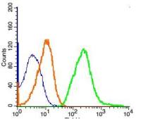
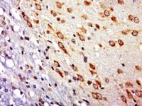
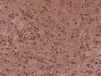
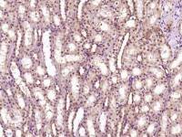
| 产品图片 | 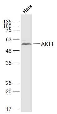 Sample: Hela(Human) Cell Lysate at 30 ug Primary: Anti-AKT1 (bs-0115R) at 1/1000 dilution Secondary: IRDye800CW Goat Anti-Rabbit IgG at 1/20000 dilution Predicted band size: 56 kD Observed band size: 56 kD 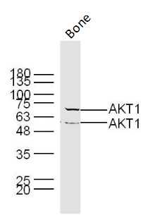 Sample: Bone (Mouse) Lysate at 40 ug Primary: Anti- AKT1 (bs- 0115R) at 1/300 dilution Secondary: IRDye800CW Goat Anti-Rabbit IgG at 1/20000 dilution Predicted band size: 56 kD Observed band size: 56/70 kD 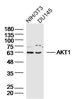 Sample: NIH/3T3 Cell (Mouse) Lysate at 40 ug DU145 Cell (Human) Lysate at 40 ug Primary: Anti-AKT1 (bs-0115R) at 1/300 dilution Secondary: IRDye800CW Goat Anti-Rabbit IgG at 1/20000 dilution Predicted band size: 56 kD Observed band size: 60 kD 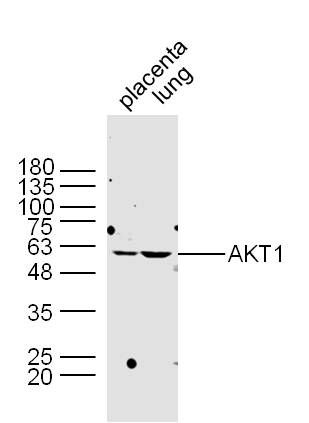 Sample: Placenta (Mouse) Lysate at 30 ug Lung (Mouse) Lysate at 30 ug Primary: Rabbit Anti-AKT1 (bs-0115R) at 1:300 dilution; Secondary: HRP conjugated Goat-Anti-rabbit IgG(bs-0295G-HRP) at 1: 5000 dilution; Predicted band size: 56 kD; Observed band size: 60 kD; 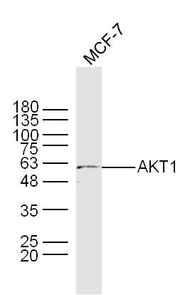 Sample: Mcf-7 Cell Lysate at 40 ug Primary: Rabbit Anti-AKT1 (bs-0115R) at 1:300 dilution; Secondary: HRP conjugated Goat-Anti-rabbit IgG(bs-0295G-HRP) at 1: 5000 dilution; Predicted band size: 56 kD; Observed band size: 60 kD; 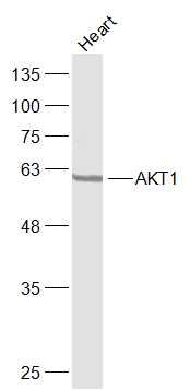 Sample: Heart(Rat) Cell Lysate at 40 ug Primary: Anti-AKT1 (bs-0115R) at 1/300 dilution Secondary: IRDye800CW Goat Anti-Rabbit IgG at 1/20000 dilution Predicted band size: 56 kD Observed band size: 63 kD 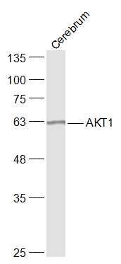 Sample: Cerebrum(Mouse) Cell Lysate at 40 ug Primary: Anti-AKT1 (bs-0115R) at 1/300 dilution Secondary: IRDye800CW Goat Anti-Rabbit IgG at 1/20000 dilution Predicted band size: 56 kD Observed band size: 63 kD 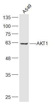 Sample: A549(Human) Cell Lysate at 30 ug Primary: Anti-AKT1 (bs-0115R) at 1/300 dilution Secondary: IRDye800CW Goat Anti-Rabbit IgG at 1/20000 dilution Predicted band size: 56 kD Observed band size: 63 kD  Sample: Spleen (Mouse) Lysate at 40 ug Primary: Anti- AKT1 (bs- 0115R) at 1/300 dilution Secondary: IRDye800CW Goat Anti-Rabbit IgG at 1/20000 dilution Predicted band size: 56 kD Observed band size: 56/70 kD 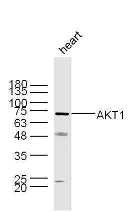 Sample: Heart (Mouse) Lysate at 40 ug Primary: Anti- AKT1 (bs- 0115R) at 1/300 dilution Secondary: IRDye800CW Goat Anti-Rabbit IgG at 1/20000 dilution Predicted band size: 56 kD Observed band size: 56/70 kD 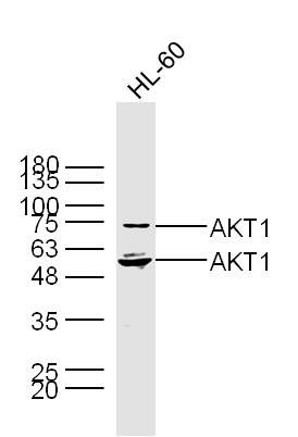 Sample: HL-60 Cell (Human) Lysate at 30 ug Primary: Anti- AKT1 (bs- 0115R) at 1/300 dilution Secondary: IRDye800CW Goat Anti-Rabbit IgG at 1/20000 dilution Predicted band size: 56 kD Observed band size: 56/70 kD 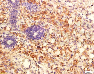 Tissue/cell: mouse transplant lymphoma; 4% Paraformaldehyde-fixed and paraffin-embedded; Antigen retrieval: citrate buffer ( 0.01M, pH 6.0 ), Boiling bathing for 15min; Block endogenous peroxidase by 3% Hydrogen peroxide for 30min; Blocking buffer (normal goat serum,C-0005) at 37℃ for 20 min; Incubation: Anti-PKB Polyclonal Antibody, Unconjugated(bs-0115R) 1:200, overnight at 4°C, followed by conjugation to the secondary antibody(SP-0023) and DAB(C-0010) staining 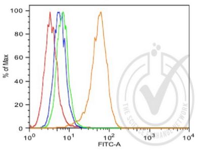 Positive control (high expression) MCF7 cells. The red histogram is unstained cells The blue histogram iscells stained with secondary antibody alone The green histogram is cells stained with rabbit IgG isotype control antibody plus secondary antibody The orange histogram is cells stained with anti-AKT1 plus secondary antibody. |
风险提示:丁香通仅作为第三方平台,为商家信息发布提供平台空间。用户咨询产品时请注意保护个人信息及财产安全,合理判断,谨慎选购商品,商家和用户对交易行为负责。对于医疗器械类产品,请先查证核实企业经营资质和医疗器械产品注册证情况。
 文献和实验
文献和实验1.留存抗体和桨细胞 抗原清除后,体内留存的抗体可特异性地直接结合再次出现的病毒颗粒、细菌和寄生虫,对后者实施中和作用和调理功能,构成记忆性抗感染的第一道防线,但发挥效应的时间不长。 抗体由浆细胞产生。浆细胞的寿命通常只有几天,称为短寿性浆细胞短寿性浆细胞系初始B细胞由抗原直接诱导产生,停留在外周淋巴器官和组织。长寿性浆细胞则经由生发中心由记忆性B细胞产生。近年来的研究揭示,此类浆细胞可长期存活。 2.记忆性B细胞 初始B细胞遭遇TD抗原后,在Th
羧肽酶B (EC 3.4.17.2) 专一用于蛋白质 C-末端碱性氨基酸(赖氨酸、精氨酸、组氨酸)的水解;又称为肽酰-L-赖氨酸(L-精氨酸)水解酶;精蛋白酶。分子量为 35kD,等电点为 6.0,最适 pH 为 7-9。羧肽酶B 是抗体碱性峰检测的特异酶,通过比较酶切前和酶切后的图谱来计算抗体碱性变体的比例,对于抗体的发酵工艺和纯化工艺稳定性的控制都具有重要的意义。 而在抗体碱性峰的检测中,没有完全统一的标准,如酶解缓冲液酶的用量,酶解时的温度和时间等。而这些与酶的性质密切
人微小病毒B19IgM抗体(B19IgM-Ab)酶联免疫分析
人 微小病毒 B19IgM 抗体( B19IgM-Ab ) 酶联免疫分析(ELISA ) 试剂盒使用说明书 本试剂仅供研究使用 目的:本试剂盒用于测定人血清,血浆及相关液体样本中 微小病毒B19IgM 抗体(B19IgM-Ab )水平 。 实验原理: 本试剂盒采用双抗原夹心酶联免疫法( ELISA )测定标本中 人 微小病毒B19IgM 抗体(B19IgM-Ab )水平 。用纯化的 人 微小
 技术资料
技术资料暂无技术资料 索取技术资料

