


- ¥1580
- 康朗生物
- kl-19881R
- 中国/美国/德国
- 2026年05月21日
- ELISA=1:500-1000 IHC-P=1:400-800 IHC-F=1:400-800 ICC=1:100-500 IF=1:100-500
- Rabbit
- Human, Cow,

企业认证
相关产品推荐更多 >

万千商家帮你免费找货
0 人在求购买到急需产品
- 详细信息
- 文献和实验
- 技术资料
- 供应商:
上海康朗生物科技有限公司
- 库存:
大量
- 目录编号:
kl-19881R
- 克隆性:
多克隆
- 抗原来源:
Rabbit
- 保质期:
12个月
- 抗体英文名:
phospho-Parkin (Ser131) antibody
- 抗体名:
磷酸化帕金森病蛋白2抗体
- 宿主:
Rabbit
- 适应物种:
Human, Cow,
- 免疫原:
KLH conjugated synthesised phosphopeptide derived from human Parkin around the phosphorylation site of Ser131.:KD(p-S)PP
- 亚型:
IgG
- 形态:
冻干粉或液体
- 应用范围:
ELISA=1:500-1000 IHC-P=1:400-800 IHC-F=1:400-800 ICC=1:100-500 IF=1:100-500
- 浓度:
1mg/ml
- 保存条件:
-20 °C
- 规格:
100ul
| 中文名称 | 磷酸化帕金森病蛋白2抗体 |
| 别 名 | Parkin (phospho S131); p-Parkin (phospho S131); AR JP; E3 ubiquitin ligase; E3 ubiquitin protein ligase parkin; E3 ubiquitin-protein ligase parkin; FRA6E; LPRS 2; LPRS2; PARK 2; PARK2; Parkin 2; Parkinson disease (autosomal recessive juvenile) 2; Parkinson disease (autosomal recessive, juvenile) 2, parkin; Parkinson disease protein 2; Parkinson juvenile disease protein 2; Parkinson protein 2 E3 ubiquitin protein ligase; Parkinson protein 2, E3 ubiquitin protein ligase (parkin); PDJ; PRKN 2; PRKN; PRKN2; PRKN2_HUMAN; Ubiquitin E3 ligase PRKN. |
| 规格价格 | 100ul/1580元 购买 大包装/询价 |
| 说 明 书 | 100ul |
| 产品类型 | 磷酸化抗体 |
| 研究领域 | 细胞生物 神经生物学 泛素 |
| 抗体来源 | Rabbit |
| 克隆类型 | Polyclonal |
| 交叉反应 | Human, Cow, |
| 产品应用 | ELISA=1:500-1000 IHC-P=1:400-800 IHC-F=1:400-800 ICC=1:100-500 IF=1:100-500 (石蜡切片需做抗原修复) not yet tested in other applications. optimal dilutions/concentrations should be determined by the end user. |
| 分 子 量 | 52kDa |
| 细胞定位 | 细胞核 细胞浆 |
| 性 状 | Lyophilized or Liquid |
| 浓 度 | 1mg/ml |
| 免 疫 原 | KLH conjugated synthesised phosphopeptide derived from human Parkin around the phosphorylation site of Ser131.:KD(p-S)PP |
| 亚 型 | IgG |
| 纯化方法 | affinity purified by Protein A |
| 储 存 液 | 0.01M TBS(pH7.4) with 1% BSA, 0.03% Proclin300 and 50% Glycerol. |
| 保存条件 | Store at -20 °C for one year. Avoid repeated freeze/thaw cycles. The lyophilized antibody is stable at room temperature for at least one month and for greater than a year when kept at -20°C. When reconstituted in sterile pH 7.4 0.01M PBS or diluent of antibody the antibody is stable for at least two weeks at 2-4 °C. |
| PubMed | PubMed |
| 产品介绍 | background: The precise function of this gene is unknown; however, the encoded protein is a component of a multiprotein E3 ubiquitin ligase complex that mediates the targeting of substrate proteins for proteasomal degradation. Mutations in this gene are known to cause Parkinson disease and autosomal recessive juvenile Parkinson disease. Alternative splicing of this gene produces multiple transcript variants encoding distinct isoforms. Additional splice variants of this gene have been described but currently lack transcript support. [provided by RefSeq, Jul 2008] Function: Functions within a multiprotein E3 ubiquitin ligase complex, catalyzing the covalent attachment of ubiquitin moieties onto substrate proteins, such as BCL2, SYT11, CCNE1, GPR37, STUB1, a 22 kDa O-linked glycosylated isoform of SNCAIP, SEPT5, ZNF746 and AIMP2. Mediates monoubiquitination as well as 'Lys-48'-linked and 'Lys-63'-linked polyubiquitination of substrates depending on the context. Participates in the removal and/or detoxification of abnormally folded or damaged protein by mediating 'Lys-63'-linked polyubiquitination of misfolded proteins such as PARK7: 'Lys-63'-linked polyubiquitinated misfolded proteins are then recognized by HDAC6, leading to their recruitment to aggresomes, followed by degradation. Mediates 'Lys-63'-linked polyubiquitination of SNCAIP, possibly playing a role in Lewy-body formation. Mediates monoubiquitination of BCL2, thereby acting as a positive regulator of autophagy. Promotes the autophagic degradation of dysfunctional depolarized mitochondria. Mediates 'Lys-48'-linked polyubiquitination of ZNF746, followed by degradation of ZNF746 by the proteasome; possibly playing a role in role in regulation of neuron death. Limits the production of reactive oxygen species (ROS). Loss of this ubiquitin ligase activity appears to be the mechanism underlying pathogenesis of PARK2. May protect neurons against alpha synuclein toxicity, proteasomal dysfunction, GPR37 accumulation, and kainate-induced excitotoxicity. May play a role in controlling neurotransmitter trafficking at the presynaptic terminal and in calcium-dependent exocytosis. Regulates cyclin-E during neuronal apoptosis. May represent a tumor suppressor gene. Subcellular Location: Cytoplasm > cytosol. Nucleus. Endoplasmic reticulum. Mitochondrion. Mainly localizes in the cytosol. Co-localizes with SYT11 in neutrites. Co-localizes with SNCAIP in brainstem Lewy bodies. Relocates to dysfunctional mitochondria that have lost the mitochondial membrane potential; recruitement to mitochondria is PINK1-dependent. Tissue Specificity: Highly expressed in the brain including the substantia nigra. Expressed in heart, testis and skeletal muscle. Expression is down-regulated or absent in tumor biopsies, and absent in the brain of PARK2 patients. Overexpression protects dopamine neurons from kainate-mediated apoptosis. Found in serum (at protein level). Post-translational modifications: Auto-ubiquitinates in an E2-dependent manner leading to its own degradation. Also polyubiquitinated by RNF41 for proteasomal degradation. S-nitrosylated. The inhibition of PARK2 ubiquitin E3 ligase activity by S-nitrosylation could contribute to the degenerative process in PD by impairing the ubiquitination of PARK2 substrates. DISEASE: Defects in PARK2 are a cause of Parkinson disease (PARK) [MIM:168600]. A complex neurodegenerative disorder characterized by bradykinesia, resting tremor, muscular rigidity and postural instability. Additional features are characteristic postural abnormalities, dysautonomia, dystonic cramps, and dementia. The pathology of Parkinson disease involves the loss of dopaminergic neurons in the substantia nigra and the presence of Lewy bodies (intraneuronal accumulations of aggregated proteins), in surviving neurons in various areas of the brain. The disease is progressive and usually manifests after the age of 50 years, although early-onset cases (before 50 years) are known. The majority of the cases are sporadic suggesting a multifactorial etiology based on environmental and genetic factors. However, some patients present with a positive family history for the disease. Familial forms of the disease usually begin at earlier ages and are associated with atypical clinical features. Defects in PARK2 are the cause of Parkinson disease type 2 (PARK2) [MIM:600116]; also known as early-onset parkinsonism with diurnal fluctuation (EPDF) or autosomal recessive juvenile Parkinson disease (PDJ). A neurodegenerative disorder characterized by bradykinesia, rigidity, postural instability, tremor, and onset usually befor 40. It differs from classic Parkinson disease by early DOPA-induced dyskinesia, diurnal fluctuation of the symptoms, sleep benefit, dystonia and hyper-reflexia. Dementia is absent. Pathologically, patients show loss of dopaminergic neurons in the substantia nigra, similar to that seen in Parkinson disease; however, Lewy bodies (intraneuronal accumulations of aggregated proteins) are absent. Note=Defects in PARK2 may be involved in the development and/or progression of ovarian cancer. Similarity: Belongs to the RBR family. Parkin subfamily. Contains 1 IBR-type zinc finger. Contains 2 RING-type zinc fingers. Contains 1 ubiquitin-like domain. SWISS: O60260 Gene ID: 5071 Database links: Entrez Gene: 5071 Human Entrez Gene: 50873 Mouse Omim: 602544 Human SwissProt: O60260 Human SwissProt: Q9WVS6 Mouse Unigene: 132954 Human Unigene: 311110 Mouse Important Note: This product as supplied is intended for research use only, not for use in human, therapeutic or diagnostic applications. |
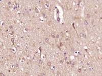
| 产品图片 | 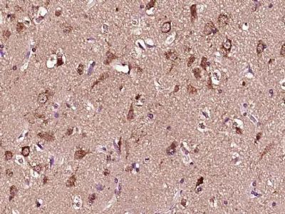 Paraformaldehyde-fixed, paraffin embedded (Human brain glioma); Antigen retrieval by boiling in sodium citrate buffer (pH6.0) for 15min; Block endogenous peroxidase by 3% hydrogen peroxide for 20 minutes; Blocking buffer (normal goat serum) at 37°C for 30min; Antibody incubation with (phospho-Parkin (Ser131) Polyclonal Antibody, Unconjugated (bs-19881R)) at 1:400 overnight at 4°C, followed by operating according to SP Kit(Rabbit) (sp-0023) instructionsand DAB staining. |
风险提示:丁香通仅作为第三方平台,为商家信息发布提供平台空间。用户咨询产品时请注意保护个人信息及财产安全,合理判断,谨慎选购商品,商家和用户对交易行为负责。对于医疗器械类产品,请先查证核实企业经营资质和医疗器械产品注册证情况。
 文献和实验
文献和实验Using Phospho‐Motif Antibodies to Determine Kinase Substrates
comprising both the phosphorylated residue and the surrounding residues that determine kinase specificity, with degenerate residues taking up the remaining positions. Currently, several categories of phospho?motif antibody are commercially available
Optimized Protocol to Make Phospho-Specific Antibodies that Work
, not simply its level of expression. In this review, we will discuss both the design of the phosphopeptide immunogen and immunization. The affinity purification of the phospho-specific antibody as well as the methods most suitable for characterizing
Absorption Control in Immunohistochemistry Using Phospho-Peptides Immobilized on Magnetic Beads
neutralization of phospho-specific antibodies with phospho-peptides immobilized on magnetic beads. This technique allows for sequestration of antibody–peptide complex from the incubation solution, minimizing the risk of formation of unblocked antibodies capable
 技术资料
技术资料暂无技术资料 索取技术资料

