


- ¥1380 - 2200
- 康朗生物
- kl-10403R
- 中国/美国/德国
- 2025年07月15日
- ELISA=1:500-1000 IHC-P=1:400-800 IHC-F=1:400-800 ICC=1:100-500 IF=1:100-500
- Rabbit
- Human, Mouse, Rat, Dog, Cow, Horse,

企业认证
相关产品推荐更多 >

万千商家帮你免费找货
0 人在求购买到急需产品
- 详细信息
- 文献和实验
- 技术资料
- 供应商:
上海康朗生物科技有限公司
- 库存:
大量
- 目录编号:
kl-10403R
- 克隆性:
多克隆
- 抗原来源:
Rabbit
- 保质期:
12个月
- 抗体英文名:
Pan Cytokeratin/p-CK antibody
- 抗体名:
广谱细胞角蛋白PCK抗体
- 宿主:
Rabbit
- 适应物种:
Human, Mouse, Rat, Dog, Cow, Horse,
- 免疫原:
KLH conjugated synthetic peptide derived from human cytokeratin 16:
- 亚型:
IgG
- 形态:
冻干粉或液体
- 应用范围:
ELISA=1:500-1000 IHC-P=1:400-800 IHC-F=1:400-800 ICC=1:100-500 IF=1:100-500
- 浓度:
1mg/ml
- 保存条件:
-20 °C
- 规格:
100ul 200ul
| 中文名称 | 广谱细胞角蛋白PCK抗体 |
| 别 名 | pan-cytokeratin; pan-CK; pan CK; P-CK; wide spectrum Cytokeratin; Cytokeratins; [cytokeratins 13, 14, 16, 17, 19, 24]. |
 |
Specific References (1) | bs-10403R has been referenced in 1 publications.
[IF=2.60] Niu YN, Wang K, Jin S, Fan DD, Wang MS, Xing NZ, Xia SJ. "The intriguing role of fibroblasts and c-Jun in the chemopreventive and therapeutic effect of finasteride on xenograft models of prostate cancer." Asian J Androl IHC-P ; Mouse.
PubMed:26698232
|
| 规格价格 | 100ul/1380元 购买 200ul/2200元 购买 大包装/询价 |
| 说 明 书 | 100ul 200ul |
| 研究领域 | |
| 抗体来源 | Rabbit |
| 克隆类型 | Polyclonal |
| 交叉反应 | Human, Mouse, Rat, Dog, Cow, Horse, |
| 产品应用 | ELISA=1:500-1000 IHC-P=1:400-800 IHC-F=1:400-800 ICC=1:100-500 IF=1:100-500 (石蜡切片需做抗原修复) not yet tested in other applications. optimal dilutions/concentrations should be determined by the end user. |
| 细胞定位 | 细胞浆 |
| 性 状 | Lyophilized or Liquid |
| 浓 度 | 1mg/ml |
| 免 疫 原 | KLH conjugated synthetic peptide derived from human cytokeratin 16: |
| 亚 型 | IgG |
| 纯化方法 | affinity purified by Protein A |
| 储 存 液 | 0.01M TBS(pH7.4) with 1% BSA, 0.03% Proclin300 and 50% Glycerol. |
| 保存条件 | Store at -20 °C for one year. Avoid repeated freeze/thaw cycles. The lyophilized antibody is stable at room temperature for at least one month and for greater than a year when kept at -20°C. When reconstituted in sterile pH 7.4 0.01M PBS or diluent of antibody the antibody is stable for at least two weeks at 2-4 °C. |
| PubMed | PubMed |
| 产品介绍 | background: Cytokeratins, a group comprising at least 29 different proteins, are characteristic of epithelial and trichocytic cells. Cytokeratins 1, 4, 5, 6, and 8 are members of the type II neutral to basic subfamily. Antibody to cytokeratins are specific markers of epithelial cell differentiation and have been widely used as tools in tumor identification and classification. Anti Pan Cytokeratin (mixture) is a broadly reactive reagent, which recognizes epitopes present in most human epithelial tissues. It facilitates typing of normal, metaplastic and neoplastic cells. Synergy between the various components results in staining amplification. This enables identification of cells, which would otherwise be stained only marginally. The mixture may aid in the discrimination of carcinomas and nonepithelial tumors such as sarcomas, lymphomas and neural tumors. It is also useful in detecting micrometastases in lymph nodes, bone marrow and other tissues and for determining the origin of poorly differentiated tumors. There are two types of cytokeratins the acidic type I cytokeratins and the basic or neutral type II cytokeratins. Cytokeratins are usually found in pairs comprising a type I cytokeratin and a type II cytokeratin. Usually the type II cytokeratins are 8kD larger than their type I counterparts. Subcellular Location: Cytoplasmic. SWISS: P13646 Gene ID: 3860 Database links: CK13: Entrez Gene: 3860 Human Omim: 148065 Human SwissProt: P13646 Human Unigene: 654550 Human CK14: Entrez Gene: 3861 Human Omim: 148066 Human SwissProt: P02533 Human Unigene: 654380 Human CK16: Entrez Gene: 3868 Human Omim: 148067 Human SwissProt: P08779 Human Unigene: 655160 Human CK17: Entrez Gene: 3872 Human Omim: 148069 Human SwissProt: Q04695 Human Unigene: 2785 Human CK19: Entrez Gene: 3880 Human Omim: 148020 Human SwissProt: P08727 Human Unigene: 654568 Human CK24: Entrez Gene: 192666 Human Omim: 607742 Human SwissProt: Q2M2I5 Human Unigene: 87383 Human Important Note: This product as supplied is intended for research use only, not for use in human, therapeutic or diagnostic applications. |
| 产品图片 |
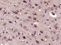
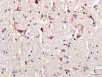
Tissue/cell: Rat brain tissue; 4% Paraformaldehyde-fixed and paraffin-embedded;
Antigen retrieval: citrate buffer ( 0.01M, pH 6.0 ), Boiling bathing for 15min; Block endogenous peroxidase by 3% Hydrogen peroxide for 30min; Blocking buffer (normal goat serum,C-0005) at 37℃ for 20 min; Incubation: Anti- Pan Cytokeratin Polyclonal Antibody, Unconjugated(bs-10403R) 1:500, overnight at 4°C, followed by conjugation to the secondary antibody(SP-0023) and DAB(C-0010) staining 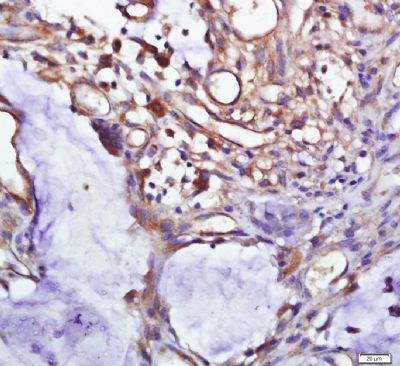
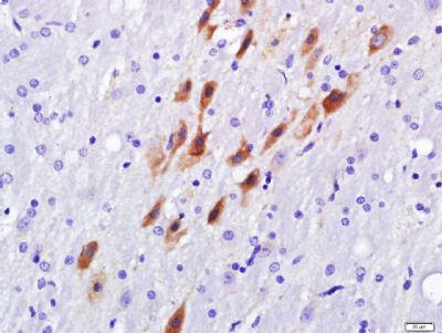
Tissue/cell: Human colon cancer; 4% Paraformaldehyde-fixed and paraffin-embedded;
Antigen retrieval: citrate buffer ( 0.01M, pH 6.0 ), Boiling bathing for 15min; Block endogenous peroxidase by 3% Hydrogen peroxide for 30min; Blocking buffer (normal goat serum,C-0005) at 37℃ for 20 min; Incubation: Anti- Pan Cytokeratin Polyclonal Antibody, Unconjugated(bs-10403R) 1:500, overnight at 4°C, followed by conjugation to the secondary antibody(SP-0023) and DAB(C-0010) staining 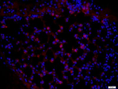
Paraformaldehyde-fixed, paraffin embedded (mouse lung); Antigen retrieval by boiling in sodium citrate buffer (pH6) for 15min; Block endogenous peroxidase by 3% hydrogen peroxide for 20 minutes; Blocking buffer (normal goat serum) at 37°C for 30min; Antibody incubation with (Pan Cytokeratin) Polyclonal Antibody, Unconjugated (bs-10403R) at 1:400 overnight at 4°C, followed by a conjugated secondary (Goat Anti-rabbit IgG/Bio) for 20minutes at 37°C, followed by a conjugated streptavidin (bs-0437P-Cy3) at[1:500] for 40 minutes and DAPI staining of the nuclei.
|
风险提示:丁香通仅作为第三方平台,为商家信息发布提供平台空间。用户咨询产品时请注意保护个人信息及财产安全,合理判断,谨慎选购商品,商家和用户对交易行为负责。对于医疗器械类产品,请先查证核实企业经营资质和医疗器械产品注册证情况。
 文献和实验
文献和实验上皮及间皮类常用标记抗体EPITHELIAL AND MESOTHELIAL MARKERSCK (MNF 116)- MNF-116 is a pan-cytokeratin antibody.- Source: Dakopatts.- Dilution 1:50.- Antigen retrieval method: HIER (pressure cooking) using EDTA.- Staining pattern: Cytoplasmic, typically
质芯片检测。此外,肽芯片或基于质谱的方法[ 20,21] 也可以用于这方面。 我们在此介绍基于蛋白质芯片技术的蛋白磷酸化筛选方法和高通量确定蛋白激酶底物的方法。我们已成功地用这种筛选工具鉴定大麦酪蛋白激酶 2α ( CK 2α) 和不同的拟南芥丝裂原活化蛋白(MAP) 激酶 [23] 的新靶标。我们这个方法使用本书第 28 章详细描述的植物蛋白质芯片(见第 28 章)。在放射性【 γ33 磷】三磷酸腺苷存在的条件下,用可溶和具有活性的激酶孵育芯片。通过磷屏成像仪或 X 射线胶片检测到的放射性信号,对可能
licanming 我的实验是用VEGF处理细胞后测定细胞PKC激活水平,拟采用Western blot的方法,用ECL法检测,这样分析PKC激活程度结果国际上认可吗? 我在Cell Signaling Technology上,查到 2261 Phospho-(Ser) PKC Substrate Antibody W IP E D 9371 Phospho-PKC (pan) (betaII Ser660) Antibody W IP
 技术资料
技术资料暂无技术资料 索取技术资料

