

- ¥780 - 2200
- 康朗生物
- kl-0681R
- 中国/美国/德国
- 2025年07月09日
- WB=1:500-2000 ELISA=1:500-1000 IHC-P=1:400-800 IHC-F=1:400-800 Flow-Cyt=1ug/Test IF=1:100-500
- Rabbit
- Human, Mouse, Rat, Chicken, Dog, Cow, Horse, Rabbit, Sheep,

企业认证
相关产品推荐更多 >

万千商家帮你免费找货
0 人在求购买到急需产品
- 详细信息
- 文献和实验
- 技术资料
- 供应商:
上海康朗生物科技有限公司
- 库存:
大量
- 目录编号:
kl-0681R
- 克隆性:
多克隆
- 抗原来源:
Rabbit
- 保质期:
12个月
- 抗体英文名:
Insulin Receptor antibody
- 抗体名:
胰岛素受体抗体
- 宿主:
Rabbit
- 适应物种:
Human, Mouse, Rat, Chicken, Dog, Cow, Horse, Rabbit, Sheep,
- 免疫原:
KLH conjugated synthetic peptide derived from human Insulin receptor subunit alpha:51-150/1384
- 亚型:
IgG
- 形态:
冻干粉或液体
- 应用范围:
WB=1:500-2000 ELISA=1:500-1000 IHC-P=1:400-800 IHC-F=1:400-800 Flow-Cyt=1ug/Test IF=1:100-500
- 浓度:
1mg/ml
- 保存条件:
-20 °C
- 规格:
50ul 100ul 200ul
| 中文名称 | 胰岛素受体抗体 |
| 别 名 | CD 220; CD220; CD220 antigen; HHF 5; HHF5; INSR; IR; INSR_HUMAN; Insulin receptor subunit alpha. |
| 规格价格 | 50ul/780元 购买 100ul/1380元 购买 200ul/2200元 购买 大包装/询价 |
| 说 明 书 | 50ul 100ul 200ul |
| 研究领域 | 细胞生物 神经生物学 信号转导 细胞凋亡 细胞膜受体 内分泌病 细胞表面分子 糖蛋白 |
| 抗体来源 | Rabbit |
| 克隆类型 | Polyclonal |
| 交叉反应 | Human, Mouse, Rat, Chicken, Dog, Cow, Horse, Rabbit, Sheep, |
| 产品应用 | WB=1:500-2000 ELISA=1:500-1000 IHC-P=1:400-800 IHC-F=1:400-800 Flow-Cyt=1ug/Test IF=1:100-500 (石蜡切片需做抗原修复) not yet tested in other applications. optimal dilutions/concentrations should be determined by the end user. |
| 分 子 量 | 80/152kDa |
| 细胞定位 | 细胞膜 |
| 性 状 | Lyophilized or Liquid |
| 浓 度 | 1mg/ml |
| 免 疫 原 | KLH conjugated synthetic peptide derived from human Insulin receptor subunit alpha:51-150/1384 |
| 亚 型 | IgG |
| 纯化方法 | affinity purified by Protein A |
| 储 存 液 | 0.01M TBS(pH7.4) with 1% BSA, 0.03% Proclin300 and 50% Glycerol. |
| 保存条件 | Store at -20 °C for one year. Avoid repeated freeze/thaw cycles. The lyophilized antibody is stable at room temperature for at least one month and for greater than a year when kept at -20°C. When reconstituted in sterile pH 7.4 0.01M PBS or diluent of antibody the antibody is stable for at least two weeks at 2-4 °C. |
| PubMed | PubMed |
| 产品介绍 | background: The insulin receptor is a heterotetrameric membrane glycoprotein with tyrosine-protein kinase activity, consisting of disulfide-linked subunits in a beta-alpha-alpha-beta configuration. The beta subunit possesses a single transmembrane domain, whereas the alpha subunit is completely extracellular. The alpha chains contribute to the formation of the ligand-binding domain, while the beta chains carry the kinase domain. Binding of insulin to the insulin receptor stimulates its association with downstream mediators including IRS1 and phosphatidylinositol 3'-kinase (PI3K) which leads to glucose uptake. Two transcript variants encoding different isoforms have been found for this gene produced by alternative splicing. Protein kinases are enzymes that transfer a phosphate group from a phosphate donor, generally the g phosphate of ATP, onto an acceptor amino acid in a substrate protein. By this basic mechanism, protein kinases mediate most of the signal transduction in eukaryotic cells, regulating cellular metabolism, transcription, cell cycle progression, cytoskeletal rearrangement and cell movement, apoptosis, and differentiation. With more than 500 gene products, the protein kinase family is one of the largest families of proteins in eukaryotes. The family has been classified in 8 major groups based on sequence comparison of their tyrosine (PTK) or serine/threonine (STK) kinase catalytic domains. The tyrosine kinase (TK) group is mainly involved in the regulation of cell-cell interactions such as differentiation, adhesion, motility and death. There are currently about 90 TK genes sequenced, 58 are of receptor protein TK (e.g. EGFR, EPH, FGFR, PDGFR, TRK, and VEGFR families), and 32 of cytosolic TK (e.g. ABL, FAK, JAK, and SRC families). Function: Receptor tyrosine kinase which mediates the pleiotropic actions of insulin. Binding of insulin leads to phosphorylation of several intracellular substrates, including, insulin receptor substrates (IRS1, 2, 3, 4), SHC, GAB1, CBL and other signaling intermediates. Each of these phosphorylated proteins serve as docking proteins for other signaling proteins that contain Src-homology-2 domains (SH2 domain) that specifically recognize different phosphotyrosines residues, including the p85 regulatory subunit of PI3K and SHP2. Phosphorylation of IRSs proteins lead to the activation of two main signaling pathways: the PI3K-AKT/PKB pathway, which is responsible for most of the metabolic actions of insulin, and the Ras-MAPK pathway, which regulates expression of some genes and cooperates with the PI3K pathway to control cell growth and differentiation. Binding of the SH2 domains of PI3K to phosphotyrosines on IRS1 leads to the activation of PI3K and the generation of phosphatidylinositol-(3, 4, 5)-triphosphate (PIP3), a lipid second messenger, which activates several PIP3-dependent serine/threonine kinases, such as PDPK1 and subsequently AKT/PKB. The net effect of this pathway is to produce a translocation of the glucose transporter SLC2A4/GLUT4 from cytoplasmic vesicles to the cell membrane to facilitate glucose transport. Moreover, upon insulin stimulation, activated AKT/PKB is responsible for: anti-apoptotic effect of insulin by inducing phosphorylation of BAD; regulates the expression of gluconeogenic and lipogenic enzymes by controlling the activity of the winged helix or forkhead (FOX) class of transcription factors. Another pathway regulated by PI3K-AKT/PKB activation is mTORC1 signaling pathway which regulates cell growth and metabolism and integrates signals from insulin. AKT mediates insulin-stimulated protein synthesis by phosphorylating TSC2 thereby activating mTORC1 pathway. The Ras/RAF/MAP2K/MAPK pathway is mainly involved in mediating cell growth, survival and cellular differentiation of insulin. Phosphorylated IRS1 recruits GRB2/SOS complex, which triggers the activation of the Ras/RAF/MAP2K/MAPK pathway. In addition to binding insulin, the insulin receptor can bind insulin-like growth factors (IGFI and IGFII). Isoform Short has a higher affinity for IGFII binding. When present in a hybrid receptor with IGF1R, binds IGF1. PubMed:12138094 shows that hybrid receptors composed of IGF1R and INSR isoform Long are activated with a high affinity by IGF1, with low affinity by IGF2 and not significantly activated by insulin, and that hybrid receptors composed of IGF1R and INSR isoform Short are activated by IGF1, IGF2 and insulin. In contrast, PubMed:16831875 shows that hybrid receptors composed of IGF1R and INSR isoform Long and hybrid receptors composed of IGF1R and INSR isoform Short have similar binding characteristics, both bind IGF1 and have a low affinity for insulin. Subunit: Tetramer of 2 alpha and 2 beta chains linked by disulfide bonds. The alpha chains contribute to the formation of the ligand-binding domain, while the beta chains carry the kinase domain. Forms a hybrid receptor with IGF1R, the hybrid is a tetramer consisting of 1 alpha chain and 1 beta chain of INSR and 1 alpha chain and 1 beta chain of IGF1R. Interacts with SORBS1 but dissociates from it following insulin stimulation. Binds SH2B2. Activated form of INSR interacts (via Tyr-999) with the PTB/PID domains of IRS1 and SHC1. The sequences surrounding the phosphorylated NPXY motif contribute differentially to either IRS1 or SHC1 recognition. Interacts (via tyrosines in the C-terminus) with IRS2 (via PTB domain and 591-786 AA); the 591-786 would be the primary anchor of IRS2 to INSR while the PTB domain would have a stabilizing action on the interaction with INSR. Interacts with the SH2 domains of the 85 kDa regulatory subunit of PI3K (PIK3R1) in vitro, when autophosphorylated on tyrosine residues. Interacts with SOCS7. Interacts (via the phosphorylated Tyr-999), with SOCS3. Interacts (via the phosphorylated Tyr-1185, Tyr-1189, Tyr-1190) with SOCS1. Interacts with CAV2 (tyrosine-phosphorylated form); the interaction is increased with 'Tyr-27'phosphorylation of CAV2 (By similarity). Interacts with ARRB2 (By similarity). Interacts with GRB10; this interaction blocks the association between IRS1/IRS2 and INSR, significantly reduces insulin-stimulated tyrosine phosphorylation of IRS1 and IRS2 and thus decreases insulin signaling. Interacts with GRB7 (By similarity). Interacts with PDPK1. Interacts (via Tyr-1190) with GRB14 (via BPS domain); this interaction protects the tyrosines in the activation loop from dephosphorylation, but promotes dephosphorylation of Tyr-999, this results in decreased interaction with, and phosphorylation of, IRS1. Interacts (via subunit alpha) with ENPP1 (via 485-599 AA); this interaction blocks autophosphorylation. Interacts with PTPRE; this interaction is dependent of Tyr-1185, Tyr-1189 and Tyr-1190 of the INSR. Interacts with STAT5B (via SH2 domain). Interacts with PTPRF. Subcellular Location: Membrane; Single-pass type I membrane protein. Tissue Specificity: Isoform Long and isoform Short are predominantly expressed in tissue targets of insulin metabolic effects: liver, adipose tissue and skeletal muscle but are also expressed in the peripheral nerve, kidney, pulmonary alveoli, pancreatic acini, placenta vascular endothelium, fibroblasts, monocytes, granulocytes, erythrocytes and skin. Isoform Short is preferentially expressed in fetal cells such as fetal fibroblasts, muscle, liver and kidney. Found as a hybrid receptor with IGF1R in muscle, heart, kidney, adipose tissue, skeletal muscle, hepatoma, fibroblasts, spleen and placenta (at protein level). Overexpressed in several tumors, including breast, colon, lung, ovary, and thyroid carcinomas. Post-translational modifications: After being transported from the endoplasmic reticulum to the Golgi apparatus, the single glycosylated precursor is further glycosylated and then cleaved, followed by its transport to the plasma membrane. Autophosphorylated on tyrosine residues in response to insulin. Phosphorylation of Tyr-999 is required for IRS1-, SHC1-, and STAT5B-binding. Dephosphorylated by PTPRE on Tyr-999, Tyr-1185, Tyr-1189 and Tyr-1190 residues. Dephosphorylated by PTPRF. DISEASE: Defects in INSR are the cause of Rabson-Mendenhall syndrome (RMS) [MIM:262190]; also known as Mendenhall syndrome. RMS is a severe insulin resistance syndrome characterized by insulin-resistant diabetes mellitus with pineal hyperplasia and somatic abnormalities. Typical features include coarse, senile-appearing facies, dental and skin abnormalities, abdominal distension, and phallic enlargement. Inheritance is autosomal recessive. Defects in INSR are the cause of leprechaunism (LEPRCH) [MIM:246200]; also known as Donohue syndrome. Leprechaunism represents the most severe form of insulin resistance syndrome, characterized by intrauterine and postnatal growth retardation and death in early infancy. Inheritance is autosomal recessive. Defects in INSR may be associated with noninsulin-dependent diabetes mellitus (NIDDM) [MIM:125853]; also known as diabetes mellitus type 2. Defects in INSR are the cause of familial hyperinsulinemic hypoglycemia type 5 (HHF5) [MIM:609968]. Familial hyperinsulinemic hypoglycemia [MIM:256450], also referred to as congenital hyperinsulinism, nesidioblastosis, or persistent hyperinsulinemic hypoglycemia of infancy (PPHI), is the most common cause of persistent hypoglycemia in infancy and is due to defective negative feedback regulation of insulin secretion by low glucose levels. Defects in INSR are the cause of insulin-resistant diabetes mellitus with acanthosis nigricans type A (IRAN type A) [MIM:610549]. This syndrome is characterized by the association of severe insulin resistance (manifested by marked hyperinsulinemia and a failure to respond to exogenous insulin) with the skin lesion acanthosis nigricans and ovarian hyperandrogenism in adolescent female subjects. Women frequently present with hirsutism, acne, amenorrhea or oligomenorrhea, and virilization. This syndrome is different from the type B that has been demonstrated to be secondary to the presence of circulating autoantibodies against the insulin receptor. Similarity: Belongs to the protein kinase superfamily. Tyr protein kinase family. Insulin receptor subfamily. Contains 3 fibronectin type-III domains. Contains 1 protein kinase domain. SWISS: P06213 Gene ID: 3643 Database links: Entrez Gene: 3643 Human Entrez Gene: 16337 Mouse Entrez Gene: 24954 Rat Entrez Gene: 484990 Dog Omim: 147670 Human SwissProt: P06213 Human SwissProt: P15208 Mouse SwissProt: P15127 Rat Unigene: 465744 Human Unigene: 9876 Rat Important Note: This product as supplied is intended for research use only, not for use in human, therapeutic or diagnostic applications. 胰岛素受体是一个四聚体,由两个α亚基和两个β亚基通过二硫键连接。两个α亚基位于细胞质膜的外侧,其上有胰岛素的结合位点;两个β亚基是跨膜蛋白,起信号转导作用。无胰岛素结 合时,受体的酪氨酸蛋白激酶没有活性。当胰岛素与受体的α亚基结合并改变了β亚基的构型后,酪氨酸蛋白激酶才被激活,激活后可催化两个反应: ①使四聚体复合物中β亚基特异位点的酪氨酸残基磷酸化,这种过程称为自我磷酸化(autophosphorylation); ②将胰岛素受体底物(insulin receptor substrate,IRSs)上具有重要作用的十几个酪氨酸残基磷酸化,磷酸化的IRSs能够结合并激活下游效应物。 (isoform CRA-c)胰岛素受体是一种跨膜蛋白,含两个α亚基(135kda)和两个β亚基(分子量:97kda)。胰岛素受体是胰岛素得传感装置,可在细胞内和细胞膜循环。 |
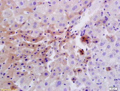
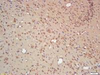
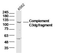
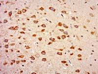
| 产品图片 | 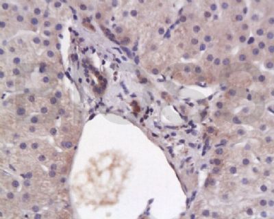 Tissue/cell: human liver tissue; 4% Paraformaldehyde-fixed and paraffin-embedded; Antigen retrieval: citrate buffer ( 0.01M, pH 6.0 ), Boiling bathing for 15min; Block endogenous peroxidase by 3% Hydrogen peroxide for 30min; Blocking buffer (normal goat serum,C-0005) at 37℃ for 20 min; Incubation: Anti-Insulin Receptor/CD220 Polyclonal Antibody, Unconjugated(bs-0681R) 1:200, overnight at 4°C, followed by conjugation to the secondary antibody(SP-0023) and DAB(C-0010) staining  Tissue/cell: rat liver tissue; 4% Paraformaldehyde-fixed and paraffin-embedded; Antigen retrieval: citrate buffer ( 0.01M, pH 6.0 ), Boiling bathing for 15min; Block endogenous peroxidase by 3% Hydrogen peroxide for 30min; Blocking buffer (normal goat serum,C-0005) at 37℃ for 20 min; Incubation: Anti-Insulin Receptor/CD220 Polyclonal Antibody, Unconjugated(bs-0681R) 1:200, overnight at 4°C, followed by conjugation to the secondary antibody(SP-0023) and DAB(C-0010) staining 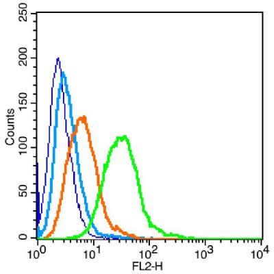 Blank control: Raji (blue). Primary Antibody: Rabbit Anti-Insulin Receptor alpha antibody(bs-0681R), Dilution: 1μg in 100 μL 1X PBS containing 0.5% BSA; Isotype Control Antibody: Rabbit IgG(orange),used under the same conditions ); Secondary Antibody: Goat anti-rabbit IgG-PE(white blue), Dilution: 1:200 in 1 X PBS containing 0.5% BSA. Protocol The cells were fixed with 2% paraformaldehyde (10 min) , then permeabilized with 90% ice-cold methanol for 30 min on ice. Primary antibody (bs-0681R, 1μg /1x10^6 cells) were incubated for 30 min on the ice, followed by 1 X PBS containing 0.5% BSA + 1 0% goat serum (15 min) to block non-specific protein-protein interactions. Then the Goat Anti-rabbit IgG/PE antibody was added into the blocking buffer mentioned above to react with the primary antibody at 1/200 dilution for 30 min on ice. Acquisition of 20,000 events was performed. |
风险提示:丁香通仅作为第三方平台,为商家信息发布提供平台空间。用户咨询产品时请注意保护个人信息及财产安全,合理判断,谨慎选购商品,商家和用户对交易行为负责。对于医疗器械类产品,请先查证核实企业经营资质和医疗器械产品注册证情况。
 文献和实验
文献和实验Assaying Tyrosine Phosphorylation of Insulin Receptor and Insulin Receptor Substrates
This chapter describes techniques to successfully detect tyrosine phosphorylation of the insulin receptor and insulin receptor substrate proteins. These assays demonstrate whether the insulin signaling pathway is activated at its earliest
胰岛素受体是一个四聚体,由两个α亚基和两个β亚基通过二硫键连接。两个α亚基位于细胞质膜的外侧,其上有胰岛素的结合位点;两个β亚基是跨膜蛋白,起信号转导作用。无胰岛素结合时,受体的酪氨酸蛋白激酶没有活性。当胰岛素与受体的α亚基结合并改变了β亚基的构型后,酪氨酸蛋白激酶才被激活,激活后可催化两个反应∶①使四聚体复合物中β亚基特异位点的酪氨酸残基磷酸化,这种过程称为自我磷酸化(autophosphorylation);②将胰岛素受体底物(insulin receptor substrate
胰岛素受体底物(insulin receptor substrate,IRSs)
蛋白质,其上有多个(至少8个)可被受体激酶磷酸化的位点,磷酸化后可同多种效应物结合,包括:PI(3)K、Syp(一种磷酸酪氨酸磷酸酶)、Nck(一种连接蛋白)、GRB2(growth factor receptor-bound protein 2,一种通过SH2同磷酸化的酪氨酸结合的连接蛋白)。第二种是Shc(是通过cDNA克隆筛选到的编码SH结构域的基因的蛋白产物),也是一种连接蛋白。Shc的酪氨酸被磷酸化后能够同GRB2结合,然后激活Ras,触发细胞的增殖。第三种底物是IRS2。IRS2的酪氨酸被磷酸
 技术资料
技术资料暂无技术资料 索取技术资料

