


- ¥780 - 2200
- 康朗生物
- kl-0395R
- 中国/美国/德国
- 2026年04月02日
- WB=1:500-2000 ELISA=1:500-1000 Flow-Cyt=1μg /test
- Rabbit
- Human, Mouse, Rat,

企业认证
相关产品推荐更多 >


万千商家帮你免费找货
0 人在求购买到急需产品
- 详细信息
- 文献和实验
- 技术资料
- 供应商:
上海康朗生物科技有限公司
- 库存:
大量
- 目录编号:
kl-0395R
- 克隆性:
多克隆
- 抗原来源:
Rabbit
- 保质期:
12个月
- 抗体英文名:
CD133 antibody
- 抗体名:
造血干细胞抗原CD133抗体
- 宿主:
Rabbit
- 适应物种:
Human, Mouse, Rat,
- 免疫原:
KLH conjugated synthetic peptide derived from human CD133:751-848/865 <Cytoplasmic>
- 亚型:
IgG
- 形态:
冻干粉或液体
- 应用范围:
WB=1:500-2000 ELISA=1:500-1000 Flow-Cyt=1μg /test
- 浓度:
1mg/ml
- 保存条件:
-20 °C
- 规格:
50ul 100ul 200ul
| 中文名称 | 造血干细胞抗原CD133抗体 |
| 别 名 | AC133; Antigen AC133; Hematopoietic stem cell antigen; hProminin; PROM1; Prominin I; Prominin like protein 1 precursor; Prominin mouse like 1; prominin1; PROML1; CD133; CORD12; MCDR2; MSTP061; PROML1; RP41; STGD4; PROM1_HUMAN; CD133 antigen. |
 |
Specific References (0) | bs-0395R has been referenced in 0 publications. |
| 规格价格 | 50ul/780元 购买 100ul/1380元 购买 200ul/2200元 购买 大包装/询价 |
| 说 明 书 | 50ul 100ul 200ul |
| 研究领域 | 肿瘤 细胞生物 免疫学 干细胞 细胞类型标志物 |
| 抗体来源 | Rabbit |
| 克隆类型 | Polyclonal |
| 交叉反应 | Human, Mouse, Rat, |
| 产品应用 | WB=1:500-2000 ELISA=1:500-1000 Flow-Cyt=1μg /test (石蜡切片需做抗原修复) not yet tested in other applications. optimal dilutions/concentrations should be determined by the end user. |
| 分 子 量 | 95kDa |
| 细胞定位 | 细胞膜 |
| 性 状 | Lyophilized or Liquid |
| 浓 度 | 1mg/ml |
| 免 疫 原 | KLH conjugated synthetic peptide derived from human CD133:751-848/865 <Cytoplasmic> |
| 亚 型 | IgG |
| 纯化方法 | affinity purified by Protein A |
| 储 存 液 | 0.01M TBS(pH7.4) with 1% BSA, 0.03% Proclin300 and 50% Glycerol. |
| 保存条件 | Store at -20 °C for one year. Avoid repeated freeze/thaw cycles. The lyophilized antibody is stable at room temperature for at least one month and for greater than a year when kept at -20°C. When reconstituted in sterile pH 7.4 0.01M PBS or diluent of antibody the antibody is stable for at least two weeks at 2-4 °C. |
| PubMed | PubMed |
| 产品介绍 | background: This gene encodes a pentaspan transmembrane glycoprotein. The protein localizes to membrane protrusions and is often expressed on adult stem cells, where it is thought to function in maintaining stem cell properties by suppressing differentiation. Mutations in this gene have been shown to result in retinitis pigmentosa and Stargardt disease. Expression of this gene is also associated with several types of cancer. This gene is expressed from at least five alternative promoters that are expressed in a tissue-dependent manner. Multiple transcript variants encoding different isoforms have been found for this gene. [provided by RefSeq, Mar 2009] Function: Binds cholesterol in cholesterol-containing plasma membrane microdomains. Proposed to play a role in apical plasma membrane organization of epithelial cells. During early retinal development acts as a key regulator of disk morphogenesis. Involved in regulation of MAPK and Akt signaling pathways. In neuroblastoma cells suppresses cell differentiation such as neurite outgrowth in a RET-dependent manner. Subunit: Interacts with CDHR1 and with actin filaments. Subcellular Location: Cell projection, cilium, photoreceptor outer segment. Isoform 1: Apical cell membrane; Multi-pass membrane protein. Cell projection, microvillus membrane; Multi-pass membrane protein. Note=Found in extracellular membrane particles in various body fluids such as cerebrospinal fluid, saliva, seminal fluid and urine. Tissue Specificity: Isoform 1 is selectively expressed on CD34 hematopoietic stem and progenitor cells in adult and fetal bone marrow, fetal liver, cord blood and adult peripheral blood. Isoform 1 is not detected on other blood cells. Isoform 1 is also expressed in a number of non-lymphoid tissues including retina, pancreas, placenta, kidney, liver, lung, brain and heart. Found in saliva within small membrane particles. Isoform 2 is predominantly expressed in fetal liver, skeletal muscle, kidney, and heart as well as adult pancreas, kidney, liver, lung, and placenta. Isoform 2 is highly expressed in fetal liver, low in bone marrow, and barely detectable in peripheral blood. Isoform 2 is expressed on hematopoietic stem cells and in epidermal basal cells (at protein level). Expressed in adult retina by rod and cone photoreceptor cells (at protein level). Post-translational modifications: Isoform 1 and isoform 2 are glycosylated. DISEASE: Defects in PROM1 are the cause of retinitis pigmentosa type 41 (RP41) [MIM:612095]; also known as retinal degeneration autosomal recessive prominin-related. RP is a retinal dystrophy belonging to the group of pigmentary retinopathies. RP is characterized by retinal pigment deposits visible on fundus examination and primary loss of rod photoreceptor cells followed by secondary loss of cone photoreceptors. Patients typically have night vision blindness and loss of midperipheral visual field. As their condition progresses, they lose their far peripheral visual field and eventually central vision as well. Defects in PROM1 are the cause of cone-rod dystrophy type 12 (CORD12) [MIM:612657]. CORD12 is an inherited retinal dystrophy characterized by retinal pigment deposits visible on fundus examination, predominantly in the macular region, and initial loss of cone photoreceptors followed by rod degeneration. This leads to decreased visual acuity and sensitivity in the central visual field, followed by loss of peripheral vision. Severe loss of vision occurs earlier than in retinitis pigmentosa. Defects in PROM1 are the cause of Stargardt disease type 4 (STGD4) [MIM:603786]. Stargardt disease is the most common hereditary macular degeneration. It is characterized by decreased central vision, atrophy of the macula and underlying retinal pigment epithelium, and frequent presence of prominent flecks in the posterior pole of the retina. Defects in PROM1 are the cause of retinal macular dystrophy type 2 (MCDR2) [MIM:608051]. MCDR2 is a bull's-eye macular dystrophy characterized by bilateral annular atrophy of retinal pigment epithelium at the macula. Similarity: Belongs to the prominin family. SWISS: O43490 Gene ID: 8842 Database links: Entrez Gene: 8842 Human Entrez Gene: 19126 Mouse Entrez Gene: 60357 Rat Omim: 604365 Human SwissProt: O43490 Human SwissProt: O54990 Mouse Unigene: 614734 Human Unigene: 6250 Mouse Unigene: 144589 Rat Important Note: This product as supplied is intended for research use only, not for use in human, therapeutic or diagnostic applications. 干细胞标志物 一般认为,VEGFR2(血管内皮生长因子受体2)是HSCs(造血干细胞)的特异性的表面标志。近来经研究发现CD133分子是HSCs(造血干细胞)特异性标志。CD133即AC133,是一个新发现的HSCs(造血干细胞)表面标志,在HSCs(造血干细胞)分化成熟过程中,CD133的含量迅速降低。EPCs(血管内皮前体细胞)区别于成熟内皮细胞的主要标志是CD133。 经研究发现内皮细胞不能结合CD133的抗体。证实分化成熟的内皮细胞不具有CD133。这些说明CD133可以作为EPCs(血管内皮前体细胞)区别于成熟内皮细胞的一个表面标志. |
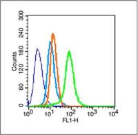
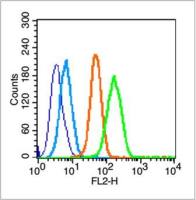
| 产品图片 |  Sample: SP2/0 (mouse)cell Lysate at 40 ug Uterus (mouse) Lysate at 40 ug Lovo (human)cell Lysate at 40 ug Primary: Anti- CD133 (bs-0395R)at 1/300 dilution Secondary: IRDye800CW Goat Anti-Rabbit IgG at 1/20000 dilution Predicted band size: 95kD Observed band size: 115 kD 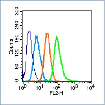 Blank control (blue line): Hep G2(blue). Primary Antibody (green line): Rabbit Anti-CD133 antibody (bs-0395R) Dilution: 1μg /10^6 cells; Isotype Control Antibody (orange line): Rabbit IgG . Secondary Antibody (white blue line): Goat anti-rabbit IgG-PE Dilution: 1μg /test. Protocol The cells were fixed with 70% ethanol Overnight at 4℃. Cells stained with Primary Antibody for 30 min at room temperature. The cells were then incubated in 1 X PBS/2%BSA/10% goat serum to block non-specific protein-protein interactions followed by the antibody for 15 min at room temperature. The secondary antibody used for 40 min at room temperature. Acquisition of 20,000 events was performed. |
风险提示:丁香通仅作为第三方平台,为商家信息发布提供平台空间。用户咨询产品时请注意保护个人信息及财产安全,合理判断,谨慎选购商品,商家和用户对交易行为负责。对于医疗器械类产品,请先查证核实企业经营资质和医疗器械产品注册证情况。
 文献和实验
文献和实验Prospective Identification of Cancer Stem Cells with the Surface Antigen CD133
Cancer cells do not share equal tumor-initiating potential. Only cancer stem cells (CSCs) can initiate cancer, which is important clinically because they should be eradicated to treat cancer patients. The purpose of experimental methods
Generation of Antibody Molecules Through Antibody Engineering
been overcome to a large extent using genetic-engineering techniques to produce chimeric mouse/human and completely human antibodies. Such an approach is particularly suitable because of the domain structure of the antibody molecule ( 2 ), where functional
The importance of antibody molecules was first recognized in the 1890s, when it was shown that immunity to tetanus and diphtheria was caused by antibodies against the bacterial exotoxins (1 ). Around the same time, it was shown that antisera
 技术资料
技术资料暂无技术资料 索取技术资料

