

- ¥1380
- 上海雅吉生物科技有限公司
- 中国
- YS-4563R
- 2025年07月16日
- WB=1:500-2000 ELISA=1:500-1000 IHC-P=1:400-800 IHC-F=1:400-800 IF=1:100-500
- Rabbit
- 见说明书

企业认证

万千商家帮你免费找货
0 人在求购买到急需产品
- 详细信息
- 文献和实验
- 技术资料
- 免疫原:
KLH conjugated
- 亚型:
IgG
- 形态:
冻干粉
- 保存条件:
Store at -20 °C for one year
- 克隆性:
多克隆
- 标记物:
见说明书
- 适应物种:
见说明书
- 保质期:
2年
- 抗原来源:
Rabbit
- 库存:
大量供应
- 供应商:
上海彩佑实业有限公司
- 宿主:
Rabbit
- 应用范围:
WB=1:500-2000 ELISA=1:500-1000 IHC-P=1:400-800 IHC-F=1:400-800 IF=1:100-500
- 浓度:
1mg/ml
- 靶点:
见说明书
- 抗体英文名:
Bcl-2
- 抗体名:
Bcl-2免疫组化荧光抗体
- 规格:
100ul
中文名称Bcl-2抗体
别 名Apoptosis regulator Bcl 2; Apoptosis regulator Bcl2; AW986256; B cell CLL/lymphoma 2; B cell leukemia/lymphoma 2; B cell lymphoma 2; Bcl 2; Bcl-2; Bcl2; BCL2 protein; C430015F12Rik; D630044D05Rik; D830018M01Rik; Leukemia/lymphoma, B-cell, 2; Oncogene B-cell leukemia 2; BCL2_HUMAN.
文献引用:
规格价格50ul/780元 购买 100ul/1380元 购买 200ul/2200元 购买 大包装/询价
说 明 书50ul 100ul 200ul
研究领域细胞生物 信号转导 细胞凋亡 细胞类型标志物 肿瘤细胞生物标志物 新陈代谢 线粒体
抗体来源Rabbit
克隆类型Polyclonal
交叉反应 Human, Mouse, Rat, Chicken, Dog, Pig, Cow, Horse, Rabbit, Guinea Pig,
产品应用WB=1:500-2000 ELISA=1:500-1000 IHC-P=1:400-800 IHC-F=1:400-800 Flow-Cyt=3ug/test IF=1:100-500 (石蜡切片需做抗原修复)
not yet tested in other applications.
optimal dilutions/concentrations should be determined by the end user.
分 子 量26kDa
细胞定位细胞核 细胞浆 细胞膜 线粒体
性 状Lyophilized or Liquid
浓 度1mg/ml
免 疫 原KLH conjugated synthetic peptide derived from human Bcl-2:101-160/236
亚 型IgG
纯化方法affinity purified by Protein A
储 存 液0.01M TBS(pH7.4) with 1% BSA, 0.03% Proclin300 and 50% Glycerol.
保存条件Store at -20 °C for one year. Avoid repeated freeze/thaw cycles. The lyophilized antibody is stable at room temperature for at least one month and for greater than a year when kept at -20°C. When reconstituted in sterile pH 7.4 0.01M PBS or diluent of antibody the antibody is stable for at least two weeks at 2-4 °C.
PubMedPubMed
产品介绍background:
The Bcl-2 gene was isolated at the chromosomal breakpoint of t(14;18)-bearing follicular B cell lymphomas(1,2).Bcl-2 blocks cell death following a variety of stimuli and confers a death-sparing effect to certain hematopoietic cell lines following growth factor withdrawal (3,5).Bcl-2 appears to function in several subcellular locations yet lacks any known motifs that would confer insight into its mechanism of action (6,7).A more recently identified protein,designated Bax p21(i.e., Bcl-associated X protein ),has extensive amino acid homology with Bcl-2 and both homodimerizes and forms heterodimers with Bcl-2(8). Overexpression of Bax accelerates apoptotic death induced by cytokine deprivation in an IL-3 dependent cell line and Bax also counters the death repressor activty of Bcl-2(8).
Function:
Suppresses apoptosis in a variety of cell systems including factor-dependent lymphohematopoietic and neural cells. Regulates cell death by controlling the mitochondrial membrane permeability. Appears to function in a feedback loop system with caspases. Inhibits caspase activity either by preventing the release of cytochrome c from the mitochondria and/or by binding to the apoptosis-activating factor (APAF-1).
Subunit:
Forms homodimers, and heterodimers with BAX, BAD, BAK and Bcl-X(L). Heterodimerization with BAX requires intact BH1 and BH2 motifs, and is necessary for anti-apoptotic activity. Interacts with EI24 (By similarity). Also interacts with APAF1, BBC3, BCL2L1, BNIPL, MRPL41 and TP53BP2. Binding to FKBP8 seems to target BCL2 to the mitochondria and probably interferes with the binding of BCL2 to its targets. Interacts with BAG1 in an ATP-dependent manner. Interacts with RAF1 (the 'Ser-338' and 'Ser-339' phosphorylated form). Interacts (via the BH4 domain) with EGLN3; the interaction prevents the formation of the BAX-BCL2 complex and inhibits the anti-apoptotic activity of BCL2. Interacts with G0S2; this interaction also prevents the formation of the anti-apoptotic BAX-BCL2 complex.
Subcellular Location:
Mitochondrion outer membrane; Single-pass membrane protein. Nucleus membrane; Single-pass membrane protein. Endoplasmic reticulum membrane; Single-pass membrane protein.
Tissue Specificity:
Expressed in a variety of tissues.
Post-translational modifications:
Phosphorylation/dephosphorylation on Ser-70 regulates anti-apoptotic activity. Growth factor-stimulated phosphorylation on Ser-70 by PKC is required for the anti-apoptosis activity and occurs during the G2/M phase of the cell cycle. In the absence of growth factors, BCL2 appears to be phosphorylated by other protein kinases such as ERKs and stress-activated kinases. Phosphorylated by MAPK8/JNK1 at Thr-69, Ser-70 and Ser-87, wich stimulates starvation-induced autophagy. Dephosphorylated by protein phosphatase 2A (PP2A).
Proteolytically cleaved by caspases during apoptosis. The cleaved protein, lacking the BH4 motif, has pro-apoptotic activity, causes the release of cytochrome c into the cytosol promoting further caspase activity.
Monoubiquitinated by PARK2, leading to increase its stability.
DISEASE:
Note=A chromosomal aberration involving BCL2 has been found in chronic lymphatic leukemia. Translocation t(14;18)(q32;q21) with immunoglobulin gene regions. BCL2 mutations found in non-Hodgkin lymphomas carrying the chromosomal translocation could be attributed to the Ig somatic hypermutation mechanism resulting in nucleotide transitions.
Similarity:
Belongs to the Bcl-2 family.
SWISS:
P49950
Gene ID:
596
Database links:
Entrez Gene: 281020 Cow
Entrez Gene: 596 Human
Entrez Gene: 12043 Mouse
Entrez Gene: 24224 Rat
Omim: 151430 Human
SwissProt: O02718 Cow
SwissProt: P10415 Human
SwissProt: P10417 Mouse
SwissProt: P49950 Rat
Unigene: 150749 Human
Unigene: 257460 Mouse
Unigene: 9996 Rat
Important Note:
This product as supplied is intended for research use only, not for use in human, therapeutic or diagnostic applications.
Bcl-2基因是指B-cell lymphoma gene。人体滤泡B细胞淋巴瘤中过量表达的原癌基因。由于染色体t(14;18)易位,将Bcl-2基因置于免疫球蛋白重链的转录调控下,使其表达失控。在细胞系中其过量表达能延长细胞存活期而不诱导细胞增殖。它是哺乳动物中细胞调亡的抑制基因。参与细胞凋亡的调控。肿瘤中的Bcl-2基因可提高侵润性瘤细胞的生存能力。主要用于滤胞型淋巴瘤、毛细管性白血病及细胞凋亡等方面的研究。
目前研究认为:Bcl-2也是细胞凋亡的一种抑制因子、参与细胞凋亡调控,可以用于各种恶性肿瘤的细胞凋亡的研究。
| 产品图片 |  Protein: Spleen(Mouse)lysate 30ug; Primary: Anti-Bcl-2(bs-0032R) at 1:300; Secondary: IRDye800CW Goat Anti-Rabbit IgG at 1/10000 dilution Predicted band size : 26kD Observed band size : 26kD |

Protein: Recombinant protein lysate 100ng;
Primary: Anti-Bcl-2(bs-0032R) at 1:200;
Secondary: HRP conjugated Goat Anti-Rabbit IgG(bs-0295G-HRP) at 1: 5000;
Predicted band size : 26kD
Observed band size : 26kD

Protein: Brain(Mouse)lysate 30ug;
Primary: Anti-Bcl-2(bs-0032R) at 1:200;
Secondary: HRP conjugated Goat Anti-Rabbit IgG(bs-0295G-HRP) at 1: 5000;
Predicted band size : 26kD
Observed band size : 26kD

Sample:Spleen (Mouse) Lysate at 40 ug
Primary: Anti-Bcl-2 (bs-0032R) at 1/300 dilution
Secondary: IRDye800CW Goat Anti-Rabbit IgG at 1/20000 dilution
Predicted band size: 26 kD
Observed band size: 26 kD

Sample:
Jurkat(Human) Cell Lysate at 30 ug
Primary: Anti-Bcl-2 (bs-0032R) at 1/300 dilution
Secondary: IRDye800CW Goat Anti-Rabbit IgG at 1/20000 dilution
Predicted band size: 26 kD
Observed band size: 26 kD

Sample:
Spleen (Mouse) Lysate at 40 ug
RAW264.7 Cell (Mouse) Lysate at 40 ug
Primary: Anti-Bcl-2 (bs-0032R) at 1/300 dilution
Secondary: IRDye800CW Goat Anti-Rabbit IgG at 1/20000 dilution
Predicted band size: 26 kD
Observed band size: 26 kD


Paraformaldehyde-fixed, paraffin embedded (Mouse brain); Antigen retrieval by boiling in sodium citrate buffer (pH6.0) for 15min; Block endogenous peroxidase by 3% hydrogen peroxide for 20 minutes; Blocking buffer (normal goat serum) at 37°C for 30min; Antibody incubation with (Bcl-2) Polyclonal Antibody, Unconjugated (bs-0032R) at 1:400 overnight at 4°C, followed by operating according to SP Kit(Rabbit) (sp-0023) instructionsand DAB staining.

Paraformaldehyde-fixed, paraffin embedded (Rat brain); Antigen retrieval by boiling in sodium citrate buffer (pH6.0) for 15min; Block endogenous peroxidase by 3% hydrogen peroxide for 20 minutes; Blocking buffer (normal goat serum) at 37°C for 30min; Antibody incubation with (Bcl-2) Polyclonal Antibody, Unconjugated (bs-0032R) at 1:400 overnight at 4°C, followed by operating according to SP Kit(Rabbit) (sp-0023) instructionsand DAB staining.

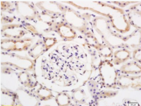
Paraformaldehyde-fixed, paraffin embedded (rat ovary tissue); Antigen retrieval by boiling in sodium citrate buffer (pH6.0) for 15min; Block endogenous peroxidase by 3% hydrogen peroxide for 20 minutes; Blocking buffer (normal goat serum) at 37°C for 30min; Antibody incubation with (Bcl-2) Polyclonal Antibody, Unconjugated (bs-0032R) at 1:400 overnight at 4°C, followed by a conjugated secondary (sp-0023) for 20 minutes and DAB staining.

Paraformaldehyde-fixed, paraffin embedded (Rat thyroid gland); Antigen retrieval by boiling in sodium citrate buffer (pH6.0) for 15min; Block endogenous peroxidase by 3% hydrogen peroxide for 20 minutes; Blocking buffer (normal goat serum) at 37°C for 30min; Antibody incubation with (B cell lymphoma 2; Bcl-2) Polyclonal Antibody, Unconjugated (bs-0032R) at 1:200 overnight at 4°C, followed by a conjugated secondary antibody (bs-0295G-cy3) for 90 minutes and DAPI for nuclei staining.
风险提示:丁香通仅作为第三方平台,为商家信息发布提供平台空间。用户咨询产品时请注意保护个人信息及财产安全,合理判断,谨慎选购商品,商家和用户对交易行为负责。对于医疗器械类产品,请先查证核实企业经营资质和医疗器械产品注册证情况。
 文献和实验
文献和实验【求助】如果要证明Bcl-2和RARP活性,应该做什么实验?
eeflying 目前有两种化合物,分别为Bcl-2和PARP的抑制剂, 虽然这两个指标通常作为细胞凋亡的指标常用于分子生物实验, 但是,如何证明这两个分子被抑制的实验却第一次碰到, 请各位战友支招,我要直接检测哪个蛋白的WB 或者做什么实验合适? magichunter 用western检测bcl-2的含量变化,检测PARP活性裂解片段的含量变化以及原型态PARP的含量
Bcl-2蛋白是bcl-2 原癌基因的编码产物,是细胞存活促进因子,属膜整合蛋白,分子量为26kDa, 定位于线粒体、内质网和连续的核周膜。Bcl-2蛋白家族是一个特别的家族,成员中有些促进凋亡,如Bad、Bid 、Bax,有些成员阻止细胞凋亡,如Bcl-2、 Bcl-x、Bcl-w。Bcl-2能够阻止细胞色素c从线粒体释放到细胞质,从而抑制了细胞凋亡。
抗体的结构决定了其对应的功能,同一抗体的V区和C区的氨基酸组成和顺序不同,决定了其功能上的差异。V区和C区的组成和结构,决定了抗体的生物学功能。 图1:抗体的主要功能(图片来源:Abbas et al: Cellular and Molecular Immunology, 7e.) 1、特异性识别功能 抗体可以通过其结构上的可变区域与抗原结合,同时,具有极高的特异性。这种特异性识别功能可以使抗体识别和结合到感染的病原体,肿瘤细胞等不同种类的分子,帮助免疫系统识别并定位到这些分子,进而启动


