


- ¥2859
- cytoskeleton
- USA
- PHDR1
- 2026年05月21日

企业认证
相关产品推荐更多 >

万千商家帮你免费找货
0 人在求购买到急需产品
- 详细信息
- 文献和实验
- 技术资料
- 英文名:
Rhodamine Phalloidin
- 供应商:
研卉生物
- 规格:
1 x 500 ul
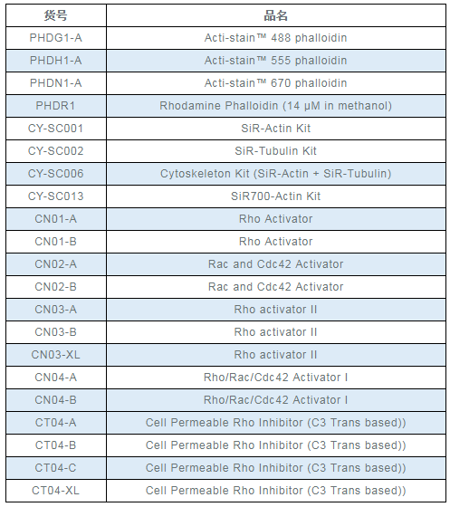
美国Cytoskeleton公司细胞骨架研究相关产品
罗丹明标记鬼笔环肽,Rhodamine Phalloidin货号:PHDR1
Product Uses Include
- Fluorescent staining of actin filaments in fixed tissue sections and tissue culture cells preparations. Note: Unlike many actin antibodies, Acti-stain™ 488 phalloidin binds only to F-actin resulting in low background fluorescence. Furthermore, binding of F-actin by Acti-stain is not appreciably different between species.
- Preparation of stabilized fluorescent actin filaments in vitro.
Actin staining is very useful in determining the structure and function of the cytoskeleton in living and fixed cells. The actin cytoskeleton is a very dynamic and labile structure in the living cell, but it can be fixed with paraformaldehyde prior to probing or staining for actin structures.
Material
Phalloidin is a seven amino acid peptide toxin from the mushroom Amanita phalloides, which binds specifically and with high affinity (Kd 20 nM) to the polymerized form of actin (F-actin). Phalloidin lowers the critical concentration of actin polymerization to less than 1 µg/ml, thereby acting as a polymerization enhancer. Phalloidin has been labeled with a proprietary green fluorescent dye which allows it to be used to stain actin filaments in tissue cultured cells and tissue sections (1, see Figure 1) and cell-free preparations. Acti-stain™ 488 phalloidin-labeled actin filaments retain many functional characteristics of unlabeled actin including their ability to interact with myosin. Actin-stain™ 488 phalloidin is supplied as an orange solid.
Note: Phalloidin is toxic and must be handled with care (LD50 human = 2mg/Kg).
Example Results and Specifications
|
|
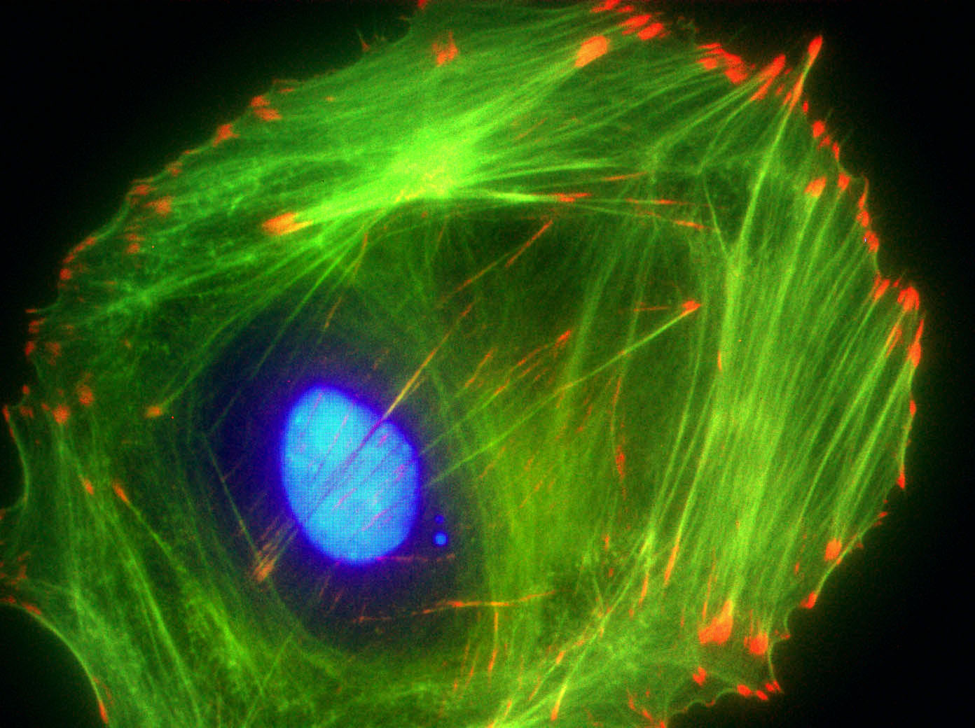
Figure 1. Actin Stress Fibers stained with Acti-stain™ 488 in a Swiss 3T3 cell. |
|
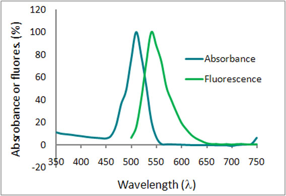
Figure 2. Emission and excitation scans for Acti-stain™ phalloidins |
|
|
|
|
|||
| Swiss 3T3 cell stained with anti-vinculin (red), Dapi (blue nucleus) and F-actin is stained with Acti-stain™ 488 (green F-actin, Cat.# PHDG1). | Absorbance and fluorescence scan of Acti-stain™ 488. Labeled phalloidin was diluted into methanol and its absorbance and excitation spectra were scanned between 350-750 and 500-750 nm, respectively. Absorbance peaks at 500 nm and fluorescence at 550 nm. |
Acti-stain phalloidins are the most well characterized phalloidins available. Tabe 1 describes their brightness, photostability, background and affinity constants to F-actin. Compare these performance characteristics to other fluorescent phalloidins and you will see the advantages of using Acti-stain™ for your actin staining requirements.
Table 1. Biochemical characteristics of fluorescent phalloidins
| Conjugate | Cat.# | Wavelengths (Ex/Em) |
Brightness (AFU*) | Stability to photobleaching(half life in seconds**) |
Background (% of total AFU at 100nM**) |
Affinity (Kd in nanomolar) |
| Fluorescein-phalloidin |
na | 485/535 FITC filter set | 432 | 6 | 22 | 72 +/-12 |
| Acti-stain™ 488 |
PHDG1 | 485/535 FITC filter set | 832 | 57 | 5 | 55 +/-8 |
| Acti-stain™ 535 |
PHDR1 | 535/585 TRITC filter set | 430 | 27 | 12 | 36 +/-7 |
| Acti-stain™ 555 |
PHDH1 | 535/585 TRITC filter set | 551 | 46 | 16 | 63 +/-8 |
| Acti-stain™ 670 |
PHDN1 | 640/680 Cy5 filter set | 332 | 8 | 18 | 50 +/-12 |
* = AFU's measured by quantitative cell imaging. ** = Measured in stained Swiss 3T3 cells in the absence of antifade.
References
1. Wulf, E. et al. (1979). Proc Natl Acad Sci USA. 76(9):4498-4502.
2. Kron, S.J. et al. (1991). Meth. Enzmol. 196: 399-416
Roitbak et al., 2011. The role of microRNAs in neural stem cell supported endothelial morphogenesis. Vascular Cell. v 3, p 25.
Yuan et al., 2012. Subsecond absolute quantitation of amine metabolites using isobaric tags for discovery of pathway activation in mammalian cells. Anal. Chem. v 84, pp 2892–2899.
风险提示:丁香通仅作为第三方平台,为商家信息发布提供平台空间。用户咨询产品时请注意保护个人信息及财产安全,合理判断,谨慎选购商品,商家和用户对交易行为负责。对于医疗器械类产品,请先查证核实企业经营资质和医疗器械产品注册证情况。
 文献和实验
文献和实验四乙基罗丹明标记抗体: 1. 称取1g RB200及2g PCL5放在乳钵中于通风橱中研磨5min; 2. 加入10ml无水丙酮,不断搅拌,5min后过滤,用滤液进行标记抗体。剩余部分吸附在滤纸上,于4℃干燥保存; 3. 量取一定量的浓度为20mg/ml的抗体,按1:1:1的比例分别加入等体积的生理盐水和0.5mol/L pH9.0的碳酸盐缓冲液来稀释抗体; 4. 再加入0.1ml的RB200溶液,边滴加边搅拌,在0—4℃中结合12—18h; 5. 用生理盐水透析5—
Rhodamine-Phalloidin/Calcofluor Staining
Rhodamine-Phalloidin/Calcofluor Staining David Amberg Grow 50 mls of yeast to 5x10E6. Add formadehyde to the media to 4% (33 mls. of 10%). Fix in media at temperature for 10 min. Spin down cells 2-3 K for 5 min
从一种毒性菇类中分离的剧毒生物碱,它同细胞松弛素的作用相反, 只与聚合的微丝结合, 而不与肌动蛋白单体分子结合。它同聚合的微丝结合后, 抑制了微丝的解体, 因而破坏了微丝的聚合和解聚的动态平衡。
 技术资料
技术资料暂无技术资料 索取技术资料

