


- $77 - 2297
- Alomone
- 以色列
- C-300
- 2026年05月21日
- 见官方网站
- Synthetic peptide
- 见官方网站

企业认证
相关产品推荐更多 >

万千商家帮你免费找货
0 人在求购买到急需产品
相关阅读
源叶生物 十万种现货试剂速发,品质稳、品类全,实验不再等!Takara 秋促来袭,提取+反转录+定量套装仅 1200 元abinScience 体内级抗体 全年锁价 低至 ¥1200阿拉丁 22万+ 科研级产品现货速发,品质严控、科研无忧- 详细信息
- 文献和实验
- 技术资料
- 免疫原:
见官方网站
- 亚型:
见官方网站
- 形态:
液体或冻干粉
- 保存条件:
-20°C
- 克隆性:
多克隆
- 标记物:
见官方网站
- 适应物种:
见官方网站
- 保质期:
6个月
- 抗原来源:
Synthetic peptide
- 目录编号:
C-300
- 级别:
高
- 库存:
大量
- 供应商:
上海信裕生物科技有限公司
- 宿主:
Synthetic peptide
- 应用范围:
见官方网站
- 浓度:
见官方网站
- 靶点:
见官方网站
- 抗体英文名:
ω-Conotoxin GVIA
- 抗体名:
ω-Conotoxin GVIA
- 规格:
0.1 mg, 0.25 mg, 0.5 mg, 1 mg, 5 mg, 10 mg
ω-Conotoxin GVIA
SNX-124A Blocker of N-Type Ca2+ Channels
Cat #: C-300
Sizes: 0.1 mg, 0.25 mg, 0.5 mg, 1 mg, 5 mg, 10 mg
Source: Synthetic peptide
MW: 3037 Da.
Net Peptide Content: 100%
Target: N-type Ca2+ channels
CLICK HERE TO RECEIVE A 0.1 mg FREE TRIAL SAMPLE!
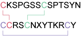
ω-Conotoxin GVIA is a synthetic toxin originally isolated from Conus geographus. ω-Conotoxin GVIA is a specific blocker of CaV2.2 Ca2+ channels. It specifically blocks N-type Cav channels by binding to the Cav2.2 α1 subunit (α1B) and its action is only partially reversible.2,3 In accordance it inhibits synaptic transmission in many systems.4 It is also reported to antagonize P2X receptors.5 The toxin is used to specifically investigate Cav2.2 channels contributions (by subtraction of the activity before and during perfusion of the toxin). Alternatively, it is used to eliminate the N-type channel contribution to highlight some other channel or enzyme activity. ω-Conotoxin GVIA was used also to purify the channel protein using immunoprecipitation techniques and to label and localize channels and synapses (See for example).6
Alomone Labs is pleased to offer ω-Conotoxin GVIA (#C-300), a highly purified synthetic peptide.
References
- Olivera, B.M. et al. (1984) Biochemistry 23 (22), 5087.
- McCleskey, E. W. et al. (1987) Proc. Natl. Acad.Sci. U.S.A. 84, 4327.
- Feng, Z. P. et al. (2001) J. Biol. Chem. 276, 15728.
- Kerr, L. M. and Yoshikami, D. (1984) Nature 308, 282.
- Lalo, U. V. et al. (2001) Brain Res. Bull. 54, 507.
- Pravettoni, E. et al. (2000) Dev. Biol. 227, 581.
For a list of product citations in the literature, see product citations tab below. If you know of additional relevant citations for this product, please let us know.
- Our Bioassay
- Specifications
- Citations
- Related Products
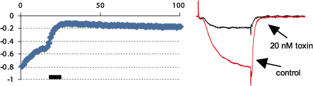
Alomone Labs ω-Conotoxin GVIA inhibits CaV2.2 heterologously expressed in Xenopus oocytes.
Left: Time course of ω-Conotoxin GVIA (#C-300) action. Current amplitudes were plotted as a function of the stimulus number. Oocyte membrane potential was held at –100 mV and 100 ms stimulating pulse to 0 mV was delivered every 10 seconds. 20 nM ω-Conotoxin GVIA were perfused in the period marked by the bar. Right: Example traces of N-type currents before and during ω-Conotoxin GVIA application.
OriginConus geographus (Geography cone).
Purity>99%.
Effective concentration20 nM - 1 μM.
SequenceCKSPGSSCSPTSYNCCRSCNXYTKRCY.
ModificationsDisulfide bonds between Cys1-Cys16, Cys8-Cys19 and Cys15-Cys26. X = Hydroxyproline. Tyr27 - C-terminal amidation.
Molecular formulaC120H188N38O43S6.
CAS number106375-28-4.
Activityω-Conotoxin GVIA specifically and reversibly blocks N-type channels1 and is reported to antagonize P2X receptors2.
Storage before reconstitutionLyophilized powder can be stored intact at room temperature for several weeks. For longer periods, it should be stored at -20°C.
ReconstitutionAny aqueous buffer. Centrifuge all product preparations before use (10000 x g 5 min).
Storage after reconstitutionUp to two weeks at 4°C or three months at -20°C.
Product citations
- Forostyak, O. et al. (2016) Stem Cell Res. 16, 622.
- Grimsley, C.A. et al. (2016) J. Neurophysiol. 116, 2550.
- Luster, B.R. et al. (2016) Physiol. Rep. 4, e12787.
- D’Onofrio, S. et al. (2016) Physiol. Rep. 4, e12740.
- Pennock, R.L. and Hentges, S.T. (2016) J. Neurophysiol. 115, 2376.
- Squecco, R. et al. (2016) Mol. Cell. Neurosci. 75, 50.
- D’Arco, M. et al. (2015) J. Neurosci. 35, 5891.
- Gan, K.J. et al. (2015) Mol. Biol. Cell. 26, 1058.
- Lopez Soto, E.J. et al. (2015) J. Gen. Physiol. 146, 205.
- Cassidy, J.S. et al. (2014) Proc. Natl. Acad. Sci. U.S.A. 111, 8979.
- Ferron, L. et al. (2014) Nat. Commun. DOI: 10.1038/ncomms4628.
- Gomez-Sanchez, R. et al. (2014) Neurobiol. Dis. 62, 426.
- Hernandez-Gonzalez, O. et al. (2014) Purinergic Signal. 10, 269.
- Perez-Burgos, A. et al. (2014) FASEB J. 28, 3064.
- Tu, H. et al. (2014) Am. J. Physiol. 306, C132.
- Alamilla, J. et al. Gillespie, D.C. (2013) PLoS ONE 8, e75688.
- Amato, A. et al. (2013) Eur. J. Pharmacol. 718, 131.
- Eggers, E.D. et al. (2013) J. Neurophysiol. 110, 709.
- Izquierdo Serra, M. et al. (2013) Biochim. Biophys. Acta 1830, 2853.
- Jones, S.L. and Stuart, G.J. (2013) J. Neurosci. 33, 19396.
- Kim, S.H. et al. (2013) J. Neurosci. 33, 8937.
- Lowe, M. et al. (2013) Exp. Physiol. 98, 1199.
- Radzicki, D. et al. (2013) J. Neurosci. 33, 9920.
- Shutov, L.P. et al. (2013) J. Physiol. 591, 2443.
- Sia, T.C. et al. (2013) Am. J. Physiol. 305, G933.
- Tzour, A. et al. (2013) J. Neuroendocrinol. 25, 76.
- Wedemeyer, C. et al. (2013) J. Neurosci. 33, 15477.
- Wen, H. et al. (2013) eLife 2, e01206.
- Wen, H. et al. (2013) J. Neurosci. 33, 7384.
- Abitbol, K. et al. (2012) J. Physiol. 590.13, 2977.
- Baillie, L.D. et al. (2012) Neuropharmacology 63, 362.
- Wang, S. et al. (2012) Cerebral Cortex 2, 584.
- Xiong, Q.J. et al. (2012) Am. J. Physiol. 303, C376.
- Alle, H. et al. (2011) J. Neurosci. 31, 8001.
- Martel, P. et al. (2011) PLoS ONE 6, e20402.
- Craviso, G.L. et al. (2010) Cell. Mol. Neurobiol. 30, 1259.
- Meunier, F.A. et al. (2010) J. Cell Sci. 123, 1131.
- Braun, M. et al. (2008) Diabetes 57, 1618.
风险提示:丁香通仅作为第三方平台,为商家信息发布提供平台空间。用户咨询产品时请注意保护个人信息及财产安全,合理判断,谨慎选购商品,商家和用户对交易行为负责。对于医疗器械类产品,请先查证核实企业经营资质和医疗器械产品注册证情况。
 文献和实验
ω -氧化 ω -oxydation 系维卡迪( P. E. Verkade)提倡的脂肪酸的氧化形式,当给动物以 C8 -C12 的脂肪酸时,从其排出二羧酸可被阐明,这些脂肪酸在羧基的相对侧的末端,即在ω的位置上。首先被氧化形成二羧酸以后,依次从两端进行β氧化。此种氧化形式在肝脏微粒体或细菌中也能见到,需要 NADPH与分子氧, P- 450和非血红素铁硫蛋白参与反应。这个反应虽不是脂肪酸氧化的主要途径,但认为部分是由ω氧化而引起的。
Measurement of [3H]PN200-110 and [125I]-Conotoxin MVIIA Binding
Intracellular Ca2+ ([Ca2+ ]i ) can rise primarily via release from intracellular storage sites (e.g., the endoplasmic reticulum) or via entry across the membrane down the steep concentration gradient (1 –3 ). Ca2+ can enter through two main
Characterization of Calcium Channel Binding
of the receptor for calcium channel blockers from skeletal muscle. Nature 328:313‐318. Wagner, J.A., Snowman, A.M., Biswas, A., Olivera, B.M., and Snyder, S.H. 1988. ω‐Conotoxin GVIA
 技术资料
暂无技术资料 索取技术资料