

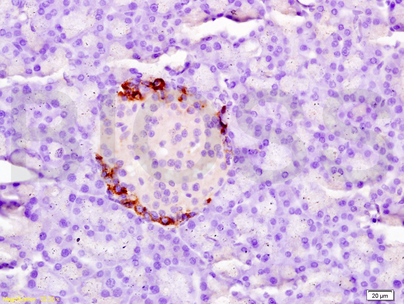
Tissue/cell: rat pancreas tissue; 4% Paraformaldehyde-fixed and paraffin-embedded;
Antigen retrieval: citrate buffer ( 0.01M, pH 6.0 ), Boiling bathing for 15min; Block endogenous peroxidase by 3% Hydrogen peroxide for 30min; Blocking buffer (normal goat serum,C-0005) at 37℃ for 20 min;
Incubation: Anti-Tissue factor/CD142/F3 Polyclonal Antibody, Unconjugated(bs-4690R) 1:200, overnight at 4°C, followed by conjugation to the secondary antibody(SP-0023) and DAB(C-0010) staining

- ¥1180 - 2800
- Bioss

- bs-4690R
- 2025年10月16日
- 产品信息以Bioss网站为准

企业认证
相关产品推荐更多 >

SNAI2 Rabbit pAb, BF750 conjugated(bs-1382R-BF750)-100ul
¥2980
FLVCR2 Rabbit pAb(bs-16147R)-50ul/100ul/200ul
¥1180
Anti-human Siglec-15 / CD33L3 (Medimmune patent Anti-Siglec-15 Biosimilar)(BIO0764SM)-1mg/5mg/20mg
¥3200
TNFSF13 Rabbit pAb, PE conjugated(bs-5725R-PE)-100ul
¥2980EGFR Rabbit pAb, Cy5.5 conjugated(bs-0405R-Cy5.5)-100ul
¥2980
万千商家帮你免费找货
0 人在求购买到急需产品
- 详细信息
- 文献和实验
- 技术资料
- 应用范围:
产品信息以Bioss网站为准
- 规格:
50ul/100ul/200ul
| 规格: | 50ul | 产品价格: | ¥1180.0 |
|---|---|---|---|
| 规格: | 100ul | 产品价格: | ¥1980.0 |
| 规格: | 200ul | 产品价格: | ¥2800.0 |
| 产品编号 | bs-4690R |
| 英文名称 | Tissue factor Rabbit pAb |
| 中文名称 | 组织因子(CD142)抗体 |
| 英文别名 | CD 142; CD142; CD142 antigen; Coagulation factor III(thromboplastin tissue factor); Coagulation factor III; F3; TF; TFA; Thromboplastin; Tissue factor; TF_HUMAN. |
| 产品应用 | WB=1:500-2000, IHC-P=1:100-500, IHC-F=1:100-500, IF=1:100-500 Not yet tested in other applications. |
| 交叉反应 | Human, Mouse, Rat (Dog, Pig, Cow, Horse, Rabbit, GuineaPig) |
| 抗体来源 | Rabbit |
| 免疫原 | KLH conjugated synthetic peptide derived from human Tissue factor |
| 亚型 | IgG |
| 性状 | Liquid |
| 纯化方法 | affinity purified by Protein A |
| 克隆类型 | Polyclonal |
| 理论分子量 | 29 kDa |
| 浓度 | 1mg/ml |
| 储存液 | 0.01M TBS (pH7.4) with 1% BSA, 0.02% Proclin300 and 50% Glycerol. |
| 研究领域 | Cardiovascular > Angiogenesis > Angiogenic Factors Cardiovascular > Blood > Coagulation > Common Cardiovascular > Blood > Coagulation > Extrinsic Kits/ Lysates/ Other > Kits > ELISA Kits > ELISA Kits > CD markers ELISA kits |
| 亚基 | Interacts with HSPE; the interaction, inhibited by heparin, promotes the generation of activated factor X and activates coagulation in the presence of activated factor VII. |
| 亚细胞定位 | Isoform 1: Membrane; Single-pass type I membrane protein.
Isoform 2: Secreted. |
| 组织特异性 | Lung, placenta and pancreas. |
| 相似性 | Belongs to the tissue factor family. |
| 功能 | Initiates blood coagulation by forming a complex with circulating factor VII or VIIa. The [TF:VIIa] complex activates factors IX or X by specific limited protolysis. TF plays a role in normal hemostasis by initiating the cell-surface assembly and propagation of the coagulation protease cascade. |
| 保存条件 | Shipped at 4℃. Store at -20℃ for one year. Avoid repeated freeze/thaw cycles. |
| 注意事项 | This product as supplied is intended for research use only, not for use in human, therapeutic or diagnostic applications. |
| 背景资料 | This gene encodes coagulation factor III which is a cell surface glycoprotein. This factor enables cells to initiate the blood coagulation cascades, and it functions as the high-affinity receptor for the coagulation factor VII. The resulting complex provides a catalytic event that is responsible for initiation of the coagulation protease cascades by specific limited proteolysis. Unlike the other cofactors of these protease cascades, which circulate as nonfunctional precursors, this factor is a potent initiator that is fully functional when expressed on cell surfaces. There are 3 distinct domains of this factor: extracellular, transmembrane, and cytoplasmic. This protein is the only one in the coagulation pathway for which a congenital deficiency has not been described. Alternate splicing results in multiple transcript variants.[provided by RefSeq, May 2010] |
| 应用 | 推荐稀释比例 |
| {WB} | {1:500-2000} |
| {IHC-P} | {1:100-500} |
| {IHC-F} | {1:100-500} |
| {IF} | {1:100-500} |
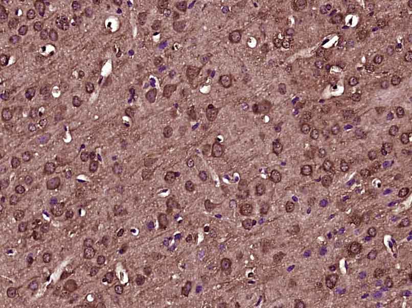
Paraformaldehyde-fixed, paraffin embedded (Mouse brain); Antigen retrieval by boiling in sodium citrate buffer (pH6.0) for 15min; Block endogenous peroxidase by 3% hydrogen peroxide for 20 minutes; Blocking buffer (normal goat serum) at 37°C for 30min; Antibody incubation with (Tissue factor) Polyclonal Antibody, Unconjugated (bs-4690R) at 1:400 overnight at 4°C, followed by operating according to SP Kit(Rabbit) (sp-0023) instructionsand DAB staining.
Sample:
Lane 1: Mouse Lung tissue lysates
Lane 2: Mouse Placenta tissue lysates
Lane 3: Human A431 cell lysates
Lane 4: Human MDA-MB-231 cell lysates
Lane 5: Human SiHa cell lysates
Primary: Anti-Tissue factor (bs-4690R) at 1/1000 dilution
Secondary: IRDye800CW Goat Anti-Rabbit IgG at 1/20000 dilution
Predicted band size: 29 kDa
Observed band size: 50 kDa
风险提示:丁香通仅作为第三方平台,为商家信息发布提供平台空间。用户咨询产品时请注意保护个人信息及财产安全,合理判断,谨慎选购商品,商家和用户对交易行为负责。对于医疗器械类产品,请先查证核实企业经营资质和医疗器械产品注册证情况。
 文献和实验
文献和实验该产品被引用文献
[IF={{ 7.13 }}] {Gleeson, Birgitta M., et al. "Bone marrow‐derived mesenchymal stem cells have innate procoagulant activity and cause microvascular obstruction following intracoronary delivery: Amelioration by anti‐thrombin therapy." STEM CELLS (2015).} {Other} {="Pig"}
[IF={{ 6.048 }}] {Takabayashi T et al. Increased expression of L‐plastin in nasal polyp of patients with nonsteroidal anti‐inflammatory drug exacerbated respiratory disease.(2018) Allergy} {IHC} {Human}
[IF={{ 5.6 }}] {Yu Muxin. et al. Neutrophil extracellular traps induce intrahepatic thrombotic tendency and liver damage in cholestatic liver disease. HEPATOL COMMUN. 2024 Aug;8(8):e0513} {IF} {Mouse}
[IF={{ 4.4 }}] {Jing-Lun Zhan. et al. Corrigendum: YKL-40 promotes chemokine expression following drug-induced liver injury via TF-PAR1 pathway in mice. FRONT PHARMACOL. 2024 Aug;15:} {WB} {Mouse}
[IF={{ 4.249 }}] {Zha C et al. Anti-β2GPI/β2GPI induces neutrophil extracellular traps formation to promote thrombogenesis via the TLR4/MyD88/MAPKs axis activation.Neuropharmacology. 2018 Aug;138:140-150.} {ICC} {Human}
相关实验
Immunostaining for Factor VIII Related Antigen (endothelium): Deparaffinize and rehydrate tissue sections. Rinse 3 times with PBS. 0.1%Pepsin in 0.1N HCL in PBS incubated sections at 37°C for 40min.
Atherosclerosis Models with Cell-Mediated Calcification
formation) at occlusion sites. These facts indicate that our model bears great similarities to clinical CTO disease. The strategy we apply here is to implant tissue-engineering scaffolds into rabbit femoral arteries and induce the cells on scaffolds
Corneal Assay for Angiogenesis
inducer (tumor tissue, cell suspension, growth factor) into a corneal pocket in order to evoke vascular outgrowth from the peripherally located limbal vasculature. In comparison to other in vivo assays, this assay has the advantage of measuring only new
 技术资料
技术资料暂无技术资料 索取技术资料
文献支持
Tissue factor Rabbit pAb(bs-4690R)-50ul/100ul/200ul
¥1180 - 2800

