


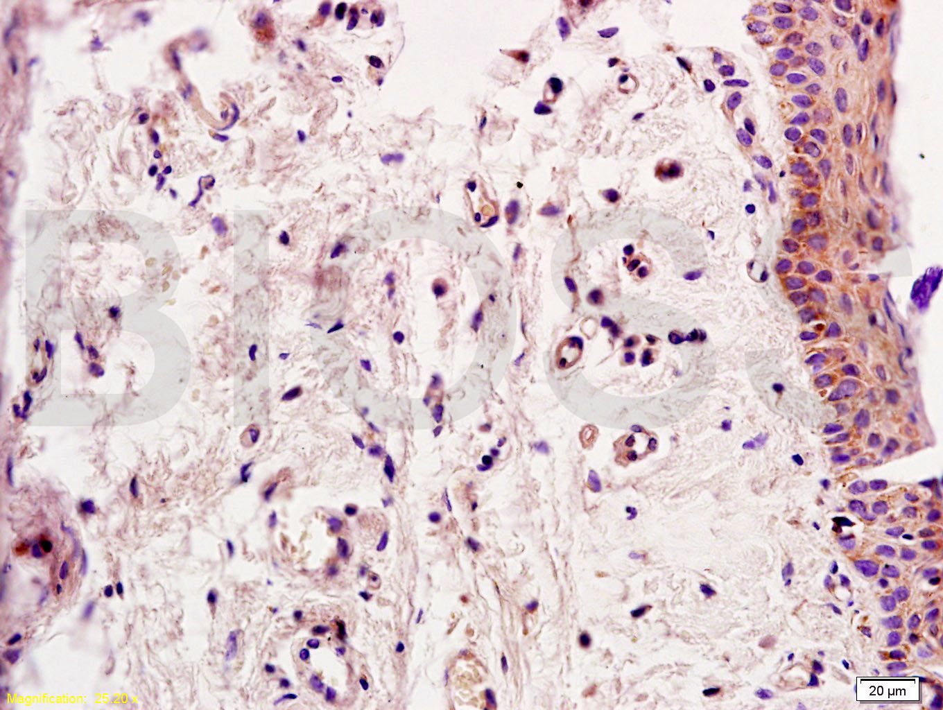
Tissue/cell: rat skin tissue; 4% Paraformaldehyde-fixed and paraffin-embedded;
Antigen retrieval: citrate buffer ( 0.01M, pH 6.0 ), Boiling bathing for 15min; Block endogenous peroxidase by 3% Hydrogen peroxide for 30min; Blocking buffer (normal goat serum,C-0005) at 37℃ for 20 min;
Incubation: Anti-Smad2 Polyclonal Antibody, Unconjugated(bs-0718R) 1:200, overnight at 4°C, followed by conjugation to the secondary antibody(SP-0023) and DAB(C-0010) staining

- ¥1180 - 2800
- Bioss

- bs-0718R
- 2025年10月24日
- 产品信息以Bioss网站为准

企业认证
相关产品推荐更多 >
万千商家帮你免费找货
0 人在求购买到急需产品
- 详细信息
- 文献和实验
- 技术资料
- 应用范围:
产品信息以Bioss网站为准
- 规格:
50ul/100ul/200ul
| 规格: | 50ul | 产品价格: | ¥1180.0 |
|---|---|---|---|
| 规格: | 100ul | 产品价格: | ¥1980.0 |
| 规格: | 200ul | 产品价格: | ¥2800.0 |
| 产品编号 | bs-0718R |
| 英文名称 | Smad2 Rabbit pAb |
| 中文名称 | 细胞信号转导分子Smad-2抗体 |
| 英文别名 | Mothers against decapentaplegic homolog 2; SMAD 2; Mothers against DPP homolog 2; Smad2; hMAD 2; hSMAD2; JV18 1; JV18; JV181; MAD; MAD Related Protein 2; MADH2; MADR2; MGC22139; MGC34440; Mothers Against Decapentaplegic Homolog 2; mothers against DPP homolog 2; SMAD 2; SMAD; SMAD2_HUMAN. |
| 产品应用 | WB=1:500-2000, IHC-P=1:100-500, IHC-F=1:100-500, IF=1:100-500, Flow-Cyt=1μg/Test Not yet tested in other applications. |
| 交叉反应 | Human, Mouse, Rat (Chicken, Dog, Cow, Rabbit) |
| 抗体来源 | Rabbit |
| 免疫原 | KLH conjugated synthetic peptide derived from human Smad2 |
| 亚型 | IgG |
| 性状 | Liquid |
| 纯化方法 | affinity purified by Protein A |
| 克隆类型 | Polyclonal |
| 理论分子量 | 52 kDa |
| 浓度 | 1mg/ml |
| 储存液 | 0.01M TBS (pH7.4) with 1% BSA, 0.02% Proclin300 and 50% Glycerol. |
| 研究领域 | Cancer > Growth factors > TGF Epigenetics and Nuclear Signaling > Nuclear Signaling Pathways > SMADs Signal Transduction > Signaling Pathway > Nuclear Signaling > SMADs Stem Cells > Signaling Pathways > TGF beta > Cytoplasmic |
| 亚基 | Momomer; the absence of TGF-beta. Heterodimer; in the presence of TGF-beta. Forms a heterodimer with co-SMAD, SMAD4, in the nucleus to form the transactivation complex SMAD2/SMAD4. Interacts with AIP1, HGS, PML and WWP1. Interacts with NEDD4L in response to TGF-beta. Found in a complex with SMAD3 and TRIM33 upon addition of TGF-beta. Interacts with ACVR1B, SMAD3 and TRIM33. Interacts (via the MH2 domain) with ZFYVE9; may form trimers with the SMAD4 co-SMAD. Interacts with FOXH1, homeobox protein TGIF, PEBP2-alpha subunit, CREB-binding protein (CBP), EP300 and SKI. Interacts with SNON; when phosphorylated at Ser-465/467. Interacts with SKOR1 and SKOR2. Interacts with PRDM16. Interacts (via MH2 domain) with LEMD3. Interacts with RBPMS. Interacts with WWP1. Interacts (dephosphorylated form, via the MH1 and MH2 domains) with RANBP3 (via its C-terminal R domain); the interaction results in the export of dephosphorylated SMAD3 out of the nucleus and termination ot the TGF-beta signaling. Interacts with PDPK1 (via PH domain). |
| 亚细胞定位 | Cytoplasm. Nucleus. Note=Cytoplasmic and nuclear in the absence of TGF-beta. On TGF-beta stimulation, migrates to the nucleus when complexed with SMAD4. On dephosphorylation by phosphatase PPM1A, released from the SMAD2/SMAD4 complex, and exported out of the nucleus by interaction with RANBP1. |
| 组织特异性 | Expressed at high levels in skeletal muscle, heart and placenta. |
| 翻译后修饰 | Phosphorylated on one or several of Thr-220, Ser-245, Ser-250, and Ser-255. In response to TGF-beta, phosphorylated on Ser-465/467 by TGF-beta and activin type 1 receptor kinases. Able to interact with SMURF2 when phosphorylated on Ser-465/467, recruiting other proteins, such as SNON, for degradation. In response to decorin, the naturally occurring inhibitor of TGF-beta signaling, phosphorylated on Ser-240 by CaMK2. Phosphorylated by MAPK3 upon EGF stimulation; which increases transcriptional activity and stability, and is blocked by calmodulin. Phosphorylated by PDPK1.
In response to TGF-beta, ubiquitinated by NEDD4L; which promotes its degradation. Acetylated on Lys-19 by coactivators in response to TGF-beta signaling, which increases transcriptional activity. Isoform short: Acetylation increases DNA binding activity in vitro and enhances its association with target promoters in vivo. Acetylation in the nucleus by EP300 is enhanced by TGF-beta. |
| 相似性 | Belongs to the dwarfin/SMAD family. Contains 1 MH1 (MAD homology 1) domain. Contains 1 MH2 (MAD homology 2) domain. |
| 功能 | Receptor-regulated SMAD (R-SMAD) that is an intracellular signal transducer and transcriptional modulator activated by TGF-beta (transforming growth factor) and activin type 1 receptor kinases. Binds the TRE element in the promoter region of many genes that are regulated by TGF-beta and, on formation of the SMAD2/SMAD4 complex, activates transcription. May act as a tumor suppressor in colorectal carcinoma. Positively regulates PDPK1 kinase activity by stimulating its dissociation from the 14-3-3 protein YWHAQ which acts as a negative regulator. |
| 保存条件 | Shipped at 4℃. Store at -20℃ for one year. Avoid repeated freeze/thaw cycles. |
| 注意事项 | This product as supplied is intended for research use only, not for use in human, therapeutic or diagnostic applications. |
| 背景资料 | The protein encoded by this gene belongs to the SMAD, a family of proteins similar to the gene products of the Drosophila gene 'mothers against decapentaplegic' (Mad) and the C. elegans gene Sma. SMAD proteins are signal transducers and transcriptional modulators that mediate multiple signaling pathways. This protein mediates the signal of the transforming growth factor (TGF)-beta, and thus regulates multiple cellular processes, such as cell proliferation, apoptosis, and differentiation. This protein is recruited to the TGF-beta receptors through its interaction with the SMAD anchor for receptor activation (SARA) protein. In response to TGF-beta signal, this protein is phosphorylated by the TGF-beta receptors. The phosphorylation induces the dissociation of this protein with SARA and the association with the family member SMAD4. The association with SMAD4 is important for the translocation of this protein into the nucleus, where it binds to target promoters and forms a transcription repressor complex with other cofactors. This protein can also be phosphorylated by activin type 1 receptor kinase, and mediates the signal from the activin. Alternatively spliced transcript variants have been observed for this gene. [provided by RefSeq, May 2012] |
| 应用 | 推荐稀释比例 |
| {WB} | {1:500-2000} |
| {IHC-P} | {1:100-500} |
| {IHC-F} | {1:100-500} |
| {IF} | {1:100-500} |
| {Flow-Cyt} | {1μg/Test} |
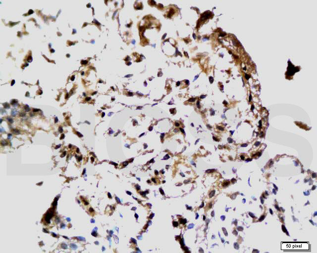
Tissue/cell: rat choroid tissue; 4% Paraformaldehyde-fixed and paraffin-embedded;
Antigen retrieval: citrate buffer ( 0.01M, pH 6.0 ), Boiling bathing for 15min; Block endogenous peroxidase by 3% Hydrogen peroxide for 30min; Blocking buffer (normal goat serum,C-0005) at 37℃ for 20 min;
Incubation: Anti-Smad2 Polyclonal Antibody, Unconjugated(bs-0718R) 1:200, overnight at 4°C, followed by conjugation to the secondary antibody(SP-0023) and DAB(C-0010) staining
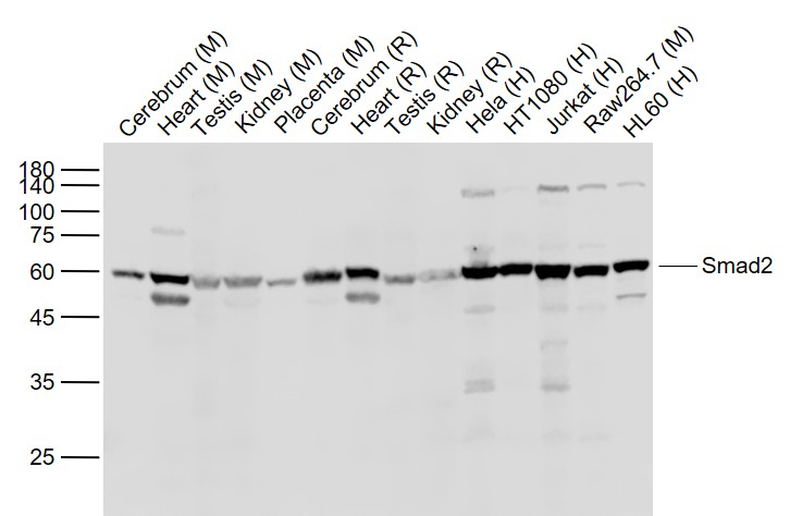
Sample:
Lane 1: Cerebrum (Mouse) Lysate at 40 ug
Lane 2: Heart (Mouse) Lysate at 40 ug
Lane 3: Testis (Mouse) Lysate at 40 ug
Lane 4: Kidney (Mouse) Lysate at 40 ug
Lane 5: Placenta (Mouse) Lysate at 40 ug
Lane 6: Cerebrum (Rat) Lysate at 40 ug
Lane 7: Heart (Rat) Lysate at 40 ug
Lane 8: Testis (Rat) Lysate at 40 ug
Lane 9: Kidney (Rat) Lysate at 40 ug
Lane 10: Hela (Human) Cell Lysate at 30 ug
Lane 11: HT1080 (Human) Cell Lysate at 30 ug
Lane 12: Jurkat (Human) Cell Lysate at 30 ug
Lane 13: Raw264.7 (Mouse) Cell Lysate at 30 ug
Lane 14: HL60 (Human) Cell Lysate at 30 ug
Primary: Anti-Smad2 (bs-0718R) at 1/1000 dilution
Secondary: IRDye800CW Goat Anti-Rabbit IgG at 1/20000 dilution
Predicted band size: 60 kD
Observed band size: 60 kD
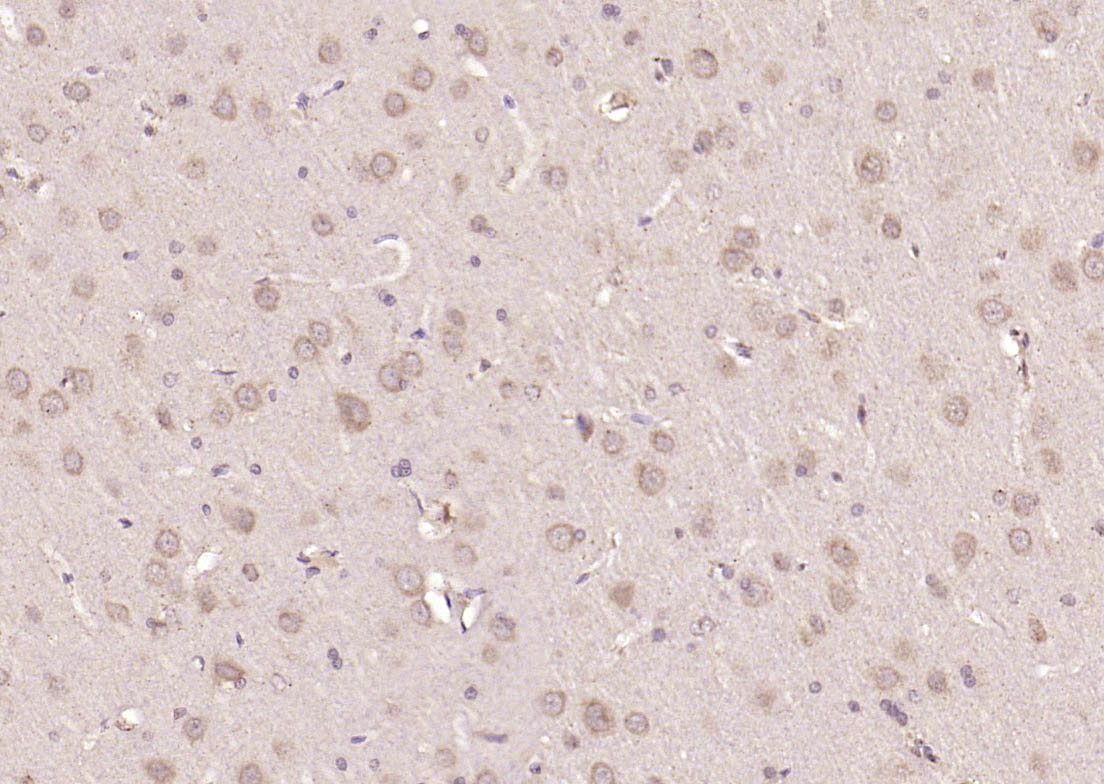
Paraformaldehyde-fixed, paraffin embedded (rat brain); Antigen retrieval by boiling in sodium citrate buffer (pH6.0) for 15min; Block endogenous peroxidase by 3% hydrogen peroxide for 20 minutes; Blocking buffer (normal goat serum) at 37°C for 30min; Antibody incubation with (Smad2) Polyclonal Antibody, Unconjugated (bs-0718R) at 1:200 overnight at 4°C, followed by operating according to SP Kit(Rabbit) (sp-0023) instructionsand DAB staining.
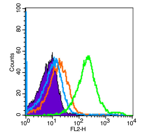
Blank control (Black line): Mouse spleen(Black).
Primary Antibody (green line): Rabbit Anti-Smad2 antibody (bs-0718R)
Dilution: 1μg /10^6 cells;
Isotype Control Antibody (orange line): Rabbit IgG .
Secondary Antibody (white blue line): Goat anti-rabbit IgG-PE
Dilution: 1μg /test.
Protocol
The cells were fixed with 4% PFA (10min at room temperature)and then permeabilized with 90% ice-cold methanol for 20 min at room temperature. The cells were then incubated in 5%BSA goat serum to block non-specific protein-protein interactions for 15 min at room temperature .Cells stained with Primary Antibody for 30 min at room temperature. The secondary antibody used for 40 min at room temperature. Acquisition of 20,000 events was performed.
风险提示:丁香通仅作为第三方平台,为商家信息发布提供平台空间。用户咨询产品时请注意保护个人信息及财产安全,合理判断,谨慎选购商品,商家和用户对交易行为负责。对于医疗器械类产品,请先查证核实企业经营资质和医疗器械产品注册证情况。
 文献和实验
文献和实验该产品被引用文献
[IF={{ 8.7 }}] {Yu Han. et al. High-precision bioactive scaffold with dECM and extracellular vesicles targeting 4E-BP inhibition for cartilage injury repair. MATER TODAY BIO. 2024 Aug;27:101114} {WB} {Rat}
[IF={{ 8.5 }}] {Genghua Chen. et al. Bulk and single-cell alternative splicing analyses reveal roles of TRA2B in myogenic differentiation. CELL PROLIFERAT. 2023 Sep;:e13545} {WB} {Chicken}
[IF={{ 7.917 }}] {Huang Shu. et al. Targeting nano-regulator based on metal–organic frameworks for enhanced immunotherapy of bone metastatic prostate cancer. CANCER NANOTECHNOL. 2023 Dec;14(1):1-15} {WB} {Mouse,Human}
[IF={{ 6.832 }}] {Li Zhang. et al. Erxian herbal pair enhances bone formation in infected bone nonunion models and attenuates lipopolysaccharide-induced osteoblastinhibition by regulating miRNA-34a-5p. BIOENGINEERED. 2022;13(6):14339-14356} {WB} {Rat}
[IF={{ 6.7 }}] {Yantong Guo. et al. Asiaticoside modulates human NK cell functional fate by mediating metabolic flexibility in the tumor microenvironment. PHYTOMEDICINE. 2024 Oct;133:155921} {WB} {Human}
相关实验
= NAL), appears to form upon oxidative cyclization of the nonfluorescent 2:1 lysine-HNE Michael adduct-Schiff base cross-link (Scheme 1). Polyclonal antibody (PAb) to the NAL-HNE fluorophore was raised in rabbit and found to be highly specific
Dynamic Monitoring of Cellular Remodeling Induced by the Transforming Growth Factor-β1
pH 7.2, 140 mM NaCl) containing 0.1% Tween 20 and 5% non-fat milk. The levels of phosphorylated (Ser465/467) and total Smad2, and expression of vimentin, a characteristic mesenchymal marker, were analyzed with specific primary antibodies (Cell
GE Healthcare Benzamidine Sepharose™ 6B is p-aminobenzamidine covalently attached to Sepharose 6B by the epoxy coupling method. p-Aminobenzamidine (PAB), is a synthetic inhibitor of trypsin-like serine protease. Trypsin and trypsin
 技术资料
技术资料暂无技术资料 索取技术资料
文献支持
Smad2 Rabbit pAb(bs-0718R)-50ul/100ul/200ul
¥1180 - 2800

