
大家都在搜

手机验证

询价列表

暂时没有已询价产品

CellTiter-Glo®
3D Cell Viability Assay
企业认证
低温保存
详询
CellTiter-Glo® 3D Cell Viability Assay
大量
北京百奥创新科技有限公司
详询
具体价格请联系我们
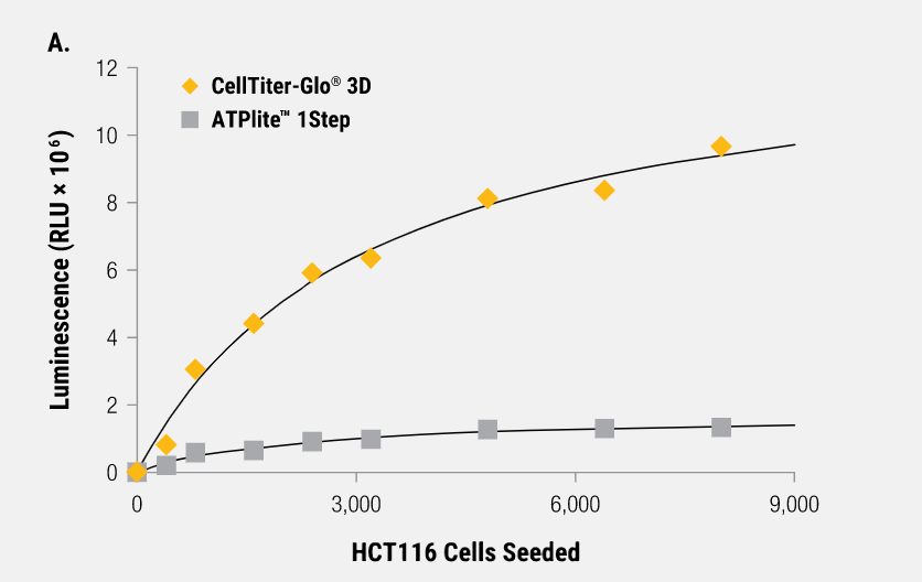
The CellTiter-Glo® 3D Cell Viability Assay is designed for determining cell viability in 3D microtissue spheroids. The assay reagent penetrates large spheroids and has increased lytic capacity—allowing more accurate determination of viability compared to other assay methods.
Based on the same reliable chemistry as the classic CellTiter-Glo® Assay, this new 3D assay reagent measures ATP as an indicator of viability and generates a luminescent readout that is much more sensitive than colorimetric or fluorescence-based methods. The simple, 30-minute protocol and ready-to-use reagent allows for fast results.
A Cell Viability Assay Validated for 3D Microtissue Cultures


风险提示:丁香通仅作为第三方平台,为商家信息发布提供平台空间。用户咨询产品时请注意保护个人信息及财产安全,合理判断,谨慎选购商品,商家和用户对交易行为负责。对于医疗器械类产品,请先查证核实企业经营资质和医疗器械产品注册证情况。
 询价记录
询价记录
 技术资料
技术资料


