


- ¥5600
- GeneTex

- 美国
- GTX60996
- 2025年07月15日
- WB, ICC/IF, IHC-Fr
- Mouse
- Human, Mouse, Rat, Bovine, Pig, Horse

企业认证
相关产品推荐更多 >
![Noggin antibody [4C12]](https://img1.dxycdn.com/2022/0328/890/5173489924628500453-14.jpg!wh200)
![PFKM antibody [AT2F11]](https://img1.dxycdn.com/2022/0328/658/4162876335210500453-14.jpg!wh200)
![Hsp90 beta antibody [3B9-D4-G4]](https://img1.dxycdn.com/2022/0328/770/3880172276057300453-14.jpg!wh200)
万千商家帮你免费找货
0 人在求购买到急需产品
- 详细信息
- 文献和实验
- 技术资料
- 免疫原:
Full length recombinant human protein expressed in and purified from E. coli.
- 亚型:
IgG1
- 形态:
Liquid
- 保存条件:
Store as concentrated solution. Centrifuge briefly prior to opening vial. For short-term storage (1-2 weeks), store at 4ºC. For long-term storage, aliquot and store at -20ºC or below. Avoid multiple freeze-thaw cycles.
- 克隆性:
Monoclonal
- 标记物:
Unconjugated
- 适应物种:
Human, Mouse, Rat, Bovine, Pig, Horse
- 保质期:
12 months from the shipping date of the product.
- 抗原来源:
Human
- 目录编号:
GTX60996
- 级别:
Primary Antibodies
- 库存:
Available
- 供应商:
GeneTex
- 宿主:
Mouse
- 应用范围:
WB, ICC/IF, IHC-Fr
- 浓度:
1 mg/ml (Please refer to the vial label for the specific concentration.)
- 靶点:
c-Fos
- 抗体英文名:
c-Fos antibody [2H2]
- 抗体名:
c-Fos 抗体 [2H2]
- 规格:
100 μl
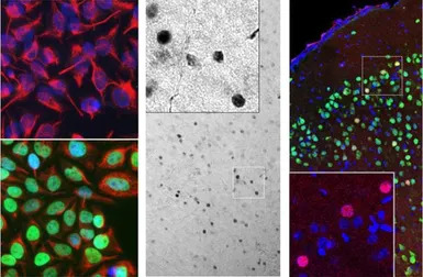
Left: GTX60996 staining (green) in HeLa cells, which were treated with serum-starvation for 36 hours, followed by 2 hours, 20% FBS stimulation (bottom panel), or PBS treatment (top panel). Green c-Fos staining only localizes in the nuclei of stimulated cells, but not in un-stimulated cells. Cells are counter-stained with our chicken polyclonal antibody against vimentin, CPCA-Vim, in red). Blue shows DAPI staining of nucleus.Middle: Mouse brain section (45 μM; fixed by transcardial perfusion with 4% PFA) labeled with GTX60996 using a standard HRP-DAB (horseradish peroxidase-3,3’-diaminobenzidine) staining technique. Cells expressing c-Fos show dark color in nucleus. Right: Mouse cortical section labeled with GTX60996 (red) and rabbit polyclonal anti Fox3/NeuN antibody (green) using immuno-fluorescent confocal-microscopy. Neurons positive for c-Fos and Fox3/NeuN appear to be yellow. Inset shows an enlarged image of GTX60996 staining. Nuclei are labeled with Dapi (blue).
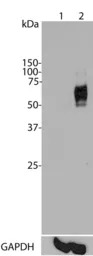
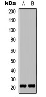
Top panel: Western blot analysis of c-Fos expression in HeLa cells using GTX60996. Lane 1: HeLa cells were serum-starved for 36 hours. 2: Serum-starved HeLa cells were stimulated with 20% FBS (fetal bovine serum) for 2 hours. GTX60996 recognizes bands in the range of 50-65 kDa, which represent multiple forms of c-Fos. Serum starvation attenuates c-Fos expression, while 20% FBS strongly stimulates c-Fos expression. Bottom panel: Blot was stripped and probed with monoclonal antibody against GAPDH.
风险提示:丁香通仅作为第三方平台,为商家信息发布提供平台空间。用户咨询产品时请注意保护个人信息及财产安全,合理判断,谨慎选购商品,商家和用户对交易行为负责。对于医疗器械类产品,请先查证核实企业经营资质和医疗器械产品注册证情况。
 文献和实验
文献和实验Chang CH et al., Cell Rep 2019 (PMID:31365864)
上海西唐生物科技有限公司 021-55229872, 65333639 www.westang.com 人C-Fos ELISA 试剂盒 原理 本实验采用双抗体夹心 ABC-ELISA 法。用抗人 C-Fos 单抗包被于酶标板上,标准品和样品中的 C-Fos与单抗结合,加入生物素化的抗人 C-Fos ,形成免疫复合物连接在板上,辣根过氧化物酶标记的 Streptavidin 与生物素结合
上海西唐生物科技有限公司 021-55229872, 65333639 www.westang.com 大鼠 C-Fos ELISA 试剂盒 原理 本实验采用双抗体夹心 ABC-ELISA 法。用抗大鼠 C-Fos 单抗包被于酶标板上,标准品和样品中的 C-Fos 与单抗结合,加入生物素化的抗大鼠 C-Fos ,形成免疫复合物连接在板上,辣根过氧化物酶标记的 Streptavidin 与生物素结合,加入底物工作液显蓝
Antibody Staining of Ovaries 1. Dissect ovaries in EBR 2. Transfer ovaries into an Eppendorf tube and add 100 µl devitellinizing buffer and 600-µl heptane 3. Agitate gently for 10 minutes
 技术资料
技术资料暂无技术资料 索取技术资料

