

- ¥9000
- GeneTex

- 美国
- GTX54824
- 2025年07月10日
- WB, ICC/IF, IHC-Fr, IP
- Rabbit
- Human, Mouse, Rat

企业认证
相关产品推荐更多 >
![CD4 (Domain 1) antibody [OX-38]](https://img1.dxycdn.com/2022/0329/854/4658064262536020453-14.jpg!wh200)

![CD13 antibody [38C12]](https://img1.dxycdn.com/2022/0328/767/1885352295197700453-14.jpg!wh200)
万千商家帮你免费找货
0 人在求购买到急需产品
- 详细信息
- 文献和实验
- 技术资料
- 免疫原:
Peptide (C)EEAGPAGEPRGSQAS, corresponding to amino acid residues 147-161 (Intracellular, N-terminus) of human HCN2 (Accession : Q9UL51).
- 亚型:
IgG
- 形态:
Liquid
- 保存条件:
Store as concentrated solution. Centrifuge briefly prior to opening vial. For short-term storage (1-2 weeks), store at 4ºC. For long-term storage, aliquot and store at -20ºC or below. Avoid multiple freeze-thaw cycles.
- 克隆性:
Polyclonal
- 标记物:
Unconjugated
- 适应物种:
Human, Mouse, Rat
- 保质期:
12 months from the shipping date of the product.
- 抗原来源:
Human
- 目录编号:
GTX54824
- 级别:
Primary Antibodies
- 库存:
Available
- 供应商:
GeneTex
- 宿主:
Rabbit
- 应用范围:
WB, ICC/IF, IHC-Fr, IP
- 浓度:
0.8 mg/ml (Please refer to the vial label for the specific concentration.)
- 靶点:
HCN2
- 抗体英文名:
HCN2 antibody
- 抗体名:
HCN2 抗体
- 规格:
50 μl
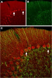
IHC-Fr analysis of rat cerebellum tissue using GTX54824 HCN2 antibody.
Panel A : HCN2 (red) appears in Purkinje cells (arrows).
Panel B : Staining of astrocytes with mouse anti-glial fibrillary acidic protein (GFAP, green) demonstrates the restriction of HCN2 to neuronal cell bodies.
Panel C : Merge of HCN2 and GFAP images demonstrates the respective localization of these proteins.

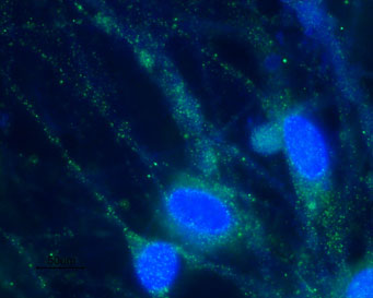
ICC/IF analysis of PFA-fixed rat DRG primary cells using GTX54824 HCN2 antibody.
Green : Primary antibody
Blue : DNA dye Hoechst 33342
Dilution : 1:100
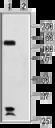
WB analysis of rat brain membrane lysate using GTX54824 HCN2 antibody preincubated with or without immunogen peptide.
Dilution : 1:200
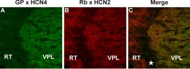
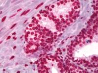
IHC-Fr analysis of mouse thalamus tissue using GTX54824 HCN2 antibody.
Panel A : Staining of HCN4 (green) appears in the ventral posterior thalamic nucleus (VPL).
Panel B : In the same section as in A, staining of HCN2 (red) appears in the ventral posterior thalamic nucleus (VPL) and also in the reticular thalamic nucleus (RT). The area between these thalamic nuclei (star) is white matter and neither protein is expressed in that region.
Panel C : Merged images of A and B.
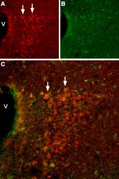
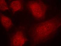
IHC-Fr analysis of mouse hypothalamus tissue using GTX54824 HCN2 antibody. (V = Third ventricle)
Panel A : HCN2 (red) appears in cells of the paraventricular nucleus (PVN, arrows).
Panel B : Staining of paraventricular nerve cells with mouse anti-calcium binding protein (CBD28k, green).
Panel C : Merge of HCN2 and CBD28k demonstrates some co-localization.
风险提示:丁香通仅作为第三方平台,为商家信息发布提供平台空间。用户咨询产品时请注意保护个人信息及财产安全,合理判断,谨慎选购商品,商家和用户对交易行为负责。对于医疗器械类产品,请先查证核实企业经营资质和医疗器械产品注册证情况。
 文献和实验
文献和实验Generation of Antibody Molecules Through Antibody Engineering
been overcome to a large extent using genetic-engineering techniques to produce chimeric mouse/human and completely human antibodies. Such an approach is particularly suitable because of the domain structure of the antibody molecule ( 2 ), where functional
The importance of antibody molecules was first recognized in the 1890s, when it was shown that immunity to tetanus and diphtheria was caused by antibodies against the bacterial exotoxins (1 ). Around the same time, it was shown that antisera
General comments: Antibodies, like most proteins, do not like to be freeze-thawed. Avoid repetitive freezing of your solution. The best way to store your antibody is to keep a high protein concentration (>1 mg/ml), add some protease
 技术资料
技术资料暂无技术资料 索取技术资料

