


- ¥4700
- GeneTex

- 美国
- GTX48819
- 2025年07月12日
- WB, IHC-P, ELISA
- Rabbit
- Human, Mouse, Rat, Bovine

企业认证
相关产品推荐更多 >
![PRRS virus M protein antibody [HL2619]](https://custom.dxycdn.com/trademd/upload/pic/2013/11/12/A1383891150.jpg!small.t.1)
![KLHL13 antibody [8D1]](https://img1.dxycdn.com/2022/0328/518/7578342753627500453-14.jpg!wh200)
![Apolipoprotein M antibody [3H3]](https://img1.dxycdn.com/2022/0328/061/2485945289388500453-14.jpg!wh200)
![Annexin A1 antibody [rANXA1/4310]](https://img1.dxycdn.com/2022/0328/763/3566284525959700453-14.jpg!wh200)
万千商家帮你免费找货
0 人在求购买到急需产品
- 详细信息
- 文献和实验
- 技术资料
- 免疫原:
This Protein A purified antibody was prepared from whole rabbit serum produced by repeated immunizations with full-length bovine S100 protein (mixture of aa homodimers and ab heterodimers).
- 亚型:
IgG
- 形态:
Liquid
- 保存条件:
Store as concentrated solution. Centrifuge briefly prior to opening vial. For short-term storage (1-2 weeks), store at 4ºC. For long-term storage, aliquot and store at -20ºC or below. Avoid multiple freeze-thaw cycles.
- 克隆性:
Polyclonal
- 标记物:
Unconjugated
- 适应物种:
Human, Mouse, Rat, Bovine
- 保质期:
12 months from the shipping date of the product.
- 抗原来源:
Bovine
- 目录编号:
GTX48819
- 级别:
Primary Antibodies
- 库存:
Available
- 供应商:
GeneTex
- 宿主:
Rabbit
- 应用范围:
WB, IHC-P, ELISA
- 浓度:
5 mg/ml (Please refer to the vial label for the specific concentration.)
- 靶点:
S100
- 抗体英文名:
S100 antibody
- 抗体名:
S100 抗体
- 规格:
100 μg
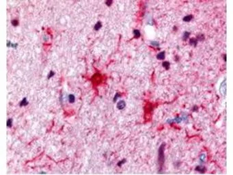
Rabbit anti-S100 was used at a 1:500 dilution to detect S100 by immunohistochemistry in human brain astrocyte tumor tissue. Tissue was formalin-fixed and paraffin embedded.
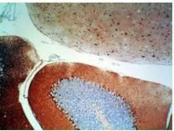
Rabbit anti-S-100 protein was used at a 1:500 dilution to detect S-100 by immunohistochemistry using a 2-step indirect method. Dark nuclear staining is observed within basket cells located near the Purkinje cells in the cerebellum. Mouse brain tissue was immersed for 24 hours in 10% neutral buffered formalin and paraffin processed followed by sectioning at 4 microns. No antigen unmasking (HIER) or protease digestion was performed prior to immunostaining. Sections were deparaffinized in xylene, and hydrated through graded alcohol to distilled water. All incubations were done at room temperature. All rinses were either distilled water or Tris-HCl with 0.05% Tween 20. Endogenous peroxidase activity was blocked with 3% Hydrogen peroxide for 10'. Primary antibody was diluted as stated and reacted for 30' followed by washes and the addition of donkey anti-rabbit HRP diluted 1:500 for 30'. DAB+ (Dakocytomation) was used as a substrate and was allowed to react for 5'.
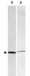
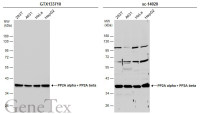
Western blot using GeneTex's Affinity Purified anti-S-100 antibody shows detection of a band ~11 kDa corresponding to bovine S-100 monomer (100 μg loaded, arrowhead lane 1). The antibody also detects S-100 from rat brain lysate (lane 2). Approximately 35 μg of a rat brain whole cell lysate was separated by 16% SDS-PAGE and transferred onto nitrocellulose. After blocking, the membrane was probed with the primary antibody diluted to 1:1,000 for 2h at room temperature followed by washes and reaction with a 1:10,000 dilution of IRDye™800 conjugated goat anti-Rabbit IgG [H&L] for 45 min at room temperature. IRDye™800 fluorescence image was captured using the Odyssey® Infrared Imaging System developed by LI-COR. IRDye is a trademark of LI-COR, Inc. Other detection systems will yield similar results.
风险提示:丁香通仅作为第三方平台,为商家信息发布提供平台空间。用户咨询产品时请注意保护个人信息及财产安全,合理判断,谨慎选购商品,商家和用户对交易行为负责。对于医疗器械类产品,请先查证核实企业经营资质和医疗器械产品注册证情况。
 文献和实验
文献和实验Yen CM et al., Neural Regen Res 2019 (PMID:31089062)
上海西唐生物科技有限公司 021-55229872, 65333639 www.westang.com 人S100 蛋白 ( S100 )ELISA 试剂盒 ( 用于血清、血浆、细胞培养上清液和其它生物体液内 ) 原理 本实验采用双抗体夹心 ABC-ELISA 法。用抗人 S100 单抗包被于酶标板上,标准品和样品中的 S100与单抗结合,加入生物素化的抗人 S100
上海西唐生物科技有限公司 021-55229872, 65333639 www.westang.com 大鼠 S100 蛋白 (S100)ELISA 试剂盒 ( 用于血清、血浆、细胞培养上清液和其它生物体液内 ) 原理 本实验采用双抗体夹心 ABC-ELISA 法。用抗大鼠 S100 单抗包被于酶标板上,标准品和样品中的 S100 与单抗结合,加入生物素化的抗大鼠 S100 ,形成免疫复合物连接
上海西唐生物科技有限公司 021-55229872, 65333639 www.westang.com 小鼠S100 蛋白 ( S100 )ELISA 试剂盒 ( 用于血清、血浆、细胞培养上清液和其它生物体液内 ) 原理 本实验采用双抗体夹心 ABC-ELISA 法。用抗小鼠 S100 单抗包被于酶标板上,标准品和样品中的 S100与单抗结合,加入生物素化的抗小鼠
 技术资料
技术资料暂无技术资料 索取技术资料

