大家都在搜

手机验证

询价列表

暂时没有已询价产品
Highly purified whole rabbit IgG
IgG
Liquid
Store as concentrated solution. Centrifuge briefly prior to opening vial. For short-term storage (1-2 weeks), store at 4ºC. For long-term storage, aliquot and store at -20ºC or below. Avoid multiple freeze-thaw cycles. Protect from light.
Polyclonal
DyLight488
Rabbit
12 months from the shipping date of the product.
Rabbit
GTX213110-04
Secondary Antibodies
Available
GeneTex
Goat
WB, ICC/IF, IHC-P, IHC-Fr, IHC-Wm, FACS
2 mg/ml (Please refer to the vial label for the specific concentration.)
Goat Anti-Rabbit IgG
Goat Anti-Rabbit IgG antibody (DyLight488)
Goat Anti-Rabbit IgG 抗体 (DyLight488)
500 μl

Double-labeled immunofluorescence photomicrographs of paraffin-embedded sections of mouse colon.
Green: E-Cadherin antibody (GTX100443) diluted at 1:500. The signal was developed using goat anti-rabbit IgG antibody (Dylight488) (GTX213110-04).
Red: alpha Tubulin antibody [GT114] (GTX628802) diluted at 1:500. The signal was developed using goat anti-mouse IgG antibody (Dylight594) (GTX213111-05).
Blue: Fluoroshield with DAPI (GTX30920).
Antigen Retrieval: Citrate buffer, pH 6.0, 15 min

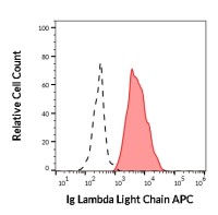
Glypican 1 antibody [N3C3] (GTX104557) detects Glypican 1 protein by flow cytometry analysis.
Sample: A431 cell.
Black: Unlabelled sample was used as a control.
Red: Glypican 1 antibody [N3C3] (GTX104557) dilution: 1:50.
Acquisition of 20,000 events were collected using the Rabbit IgG (DyLight488) (GTX113110-04) secondary antibody for FACS analysis.

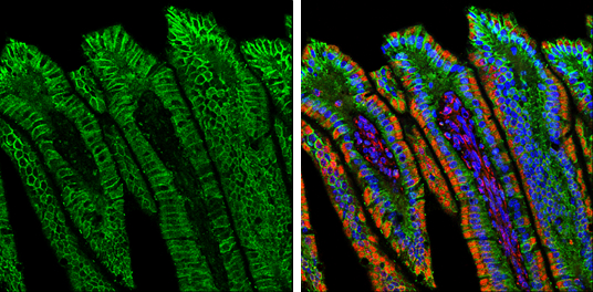
Whole mount immunohistochemical analysis of PFA-fixed 2 day-post-fertilization zebrafish embryo using Pax2a antibody (GTX128127) detected by anti-rabbit IgG antibody (Dylight488) (GTX213110-04). GTX128127 diluted at 1:100 and incubated overnight at 4ºC. GTX213110-04 diluted at 1:500 and incubated 3 hours at room temperature.

Double-labeled immunofluorescence photomicrographs of frozen sections of mouse brain.
Green: Vimentin antibody (GTX100619) diluted at 1:200. The signal was developed using goat anti-rabbit IgG antibody (Dylight488) (GTX213110-04).
Red: ME1 antibody [GT15611] (GTX632190) diluted at 1:200. The signal was developed using goat anti-mouse IgG antibody (Dylight594) (GTX213111-05).
Blue: Nuclear staining with Hoechst 33342.

alpha Tubulin antibody detects alpha Tubulin protein at cytoskeleton by immunofluorescent analysis.Sample: HeLa cells were fixed in 4% PFA at RT for 15 min.Green: alpha Tubulin stained by alpha Tubulin antibody (GTX112141) diluted at 1:500.The signal was developed using Goat Anti-Rabbit IgG antibody (DyLight488) (GTX213110-04) diluted at 1:2000.Blue: Fluoroshield with DAPI (GTX30920).

CD81 antibody (GTX101766) detects CD81 protein by flow cytometry analysis.
Sample: THP-1 cell.
Black: Unlabelled sample was used as a control.
Red: CD81 antibody (GTX101766) dilution: 1:50.
The Rabbit IgG antibody (DyLight488) (GTX213110-04) was used to detect the primary antibody.
风险提示:丁香通仅作为第三方平台,为商家信息发布提供平台空间。用户咨询产品时请注意保护个人信息及财产安全,合理判断,谨慎选购商品,商家和用户对交易行为负责。对于医疗器械类产品,请先查证核实企业经营资质和医疗器械产品注册证情况。