


- ¥1700 - 4000
- GeneTex

- 美国
- GTX128116
- 2025年07月14日
- WB, ICC/IF, IHC-P, IHC-Wm, IP, ChIP assay
- Rabbit
- Human, Mouse, Rat, Zebrafish, Bovine

企业认证
相关产品推荐更多 >
![PDGF Receptor alpha antibody [16A1] (APC)](https://img1.dxycdn.com/2022/0328/483/9364130914226500453-14.jpg!wh200)
![CD1a antibody [7A7]](https://img1.dxycdn.com/2022/0328/982/6408272936392500453-14.jpg!wh200)
![MAD2L1 antibody [17D10]](https://img1.dxycdn.com/2022/0328/985/6565065723266200453-14.jpg!wh200)
![ZEB2 antibody [OTI1E12]](https://img1.dxycdn.com/2022/0328/885/1106579086006700453-14.jpg!wh200)
万千商家帮你免费找货
0 人在求购买到急需产品
- 详细信息
- 文献和实验
- 技术资料
- 免疫原:
Carrier-protein conjugated synthetic peptide surrounding phospho Ser10 of human Histone H3. The exact sequence is proprietary.
- 亚型:
IgG
- 形态:
Liquid
- 保存条件:
Store as concentrated solution. Centrifuge briefly prior to opening vial. For short-term storage (1-2 weeks), store at 4℃. For long-term storage, aliquot and store at -20℃ or below. Avoid multiple freeze-thaw cycles.
- 克隆性:
Polyclonal
- 标记物:
Unconjugated
- 适应物种:
Human, Mouse, Rat, Zebrafish, Bovine
- 保质期:
12 months from the shipping date of the product.
- 抗原来源:
Human
- 目录编号:
GTX128116
- 级别:
Primary Antibodies
- 库存:
Available
- 供应商:
GeneTex
- 宿主:
Rabbit
- 应用范围:
WB, ICC/IF, IHC-P, IHC-Wm, IP, ChIP assay
- 浓度:
0.43 mg/ml(Please refer to the vial label for the specific concentration.)
- 靶点:
Histone H3S10ph (phospho Ser10)
- 抗体英文名:
Histone H3S10ph (phospho Ser10) antibody
- 抗体名:
Histone H3S10ph (phospho Ser10) 抗体
- 规格:
100 μl/25 μl
| 规格: | 100 μl | 产品价格: | ¥4000.0 |
|---|---|---|---|
| 规格: | 25 μl | 产品价格: | ¥1700.0 |

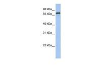
Untreated (–) and treated (+) HeLa whole cell extracts (5 μg) were separated by 15% SDS-PAGE, and the membrane was blotted with Histone H3S10ph (phospho Ser10) antibody (GTX128116) diluted at 1:2000.

Histone H3S10ph (phospho Ser10) antibody detects Histone H3S10ph (phospho Ser10) protein on zebrafish by whole mount immunohistochemical analysis.
Sample: PFA-fixed 2 day-post-fertilization zebrafish embryo.
Histone H3S10ph (phospho Ser10) (GTX128116) antibody dilution: 1:100.

Histone H3 (phospho Ser10) antibody detects HIST1H3A protein at nucleus on human gastric cancer by immunohistochemical analysis.
Sample: Paraffin-embedded gastric cancer.
Histone H3 (phospho Ser10) antibody (GTX128116) dilution: 1:500.
Antigen Retrieval: Trilogy™ (EDTA based, pH 8.0) buffer, 15min

Histone H3S10ph (phospho Ser10) antibody detects Histone H3S10ph (phospho Ser10) protein at nucleus by immunohistochemical analysis.
Sample: Paraffin-embedded mouse stomach.
Histone H3S10ph (phospho Ser10) stained by Histone H3S10ph (phospho Ser10) antibody (GTX128116) diluted at 1:500.
Antigen Retrieval: Citrate buffer, pH 6.0, 15 min

Histone H3S10ph (phospho Ser10) antibody immunoprecipitates HIST1H3B protein in IP experiments.
IP samples: 293T whole cell extract treat with 100ng/ml Nocodazole for 24 hr
A. 30 μg 293T whole cell extract treat with 100ng/ml Nocodazole for 24 hr
B. Control with 4 μg of preimmune Rabbit IgG
C. Immunoprecipitation of HIST1H3B protein by 4 μg Histone H3S10ph (phospho Ser10) antibody (GTX128116)
15 % SDS-PAGE
The immunoprecipitated HIST1H3B protein was detected by Histone H3S10ph (phospho Ser10) antibody (GTX128116) diluted at 1:1000.
[EasyBlot anti-rabbit IgG (GTX221666-01) was used as a secondary reagent]

Histone H3 (phospho Ser10) antibody detects Histone H3 (phospho Ser10) protein by western blot analysis.
A. 30 μg Rat2 whole cell lysate/extract (untreated)
B. 30 μg Rat2 whole cell lysate/extract (100 ng/ml Nocodazole treatment for 24 hr)
15% SDS-PAGE
Histone H3 (phospho Ser10) antibody (GTX128116) dilution: 1:500
The HRP-conjugated anti-rabbit IgG antibody (GTX213110-01) was used to detect the primary antibody.

Cross-linked ChIP was performed with HeLa chromatin extract treated with Nocodazole (100 ng/ml for 24 h) and 5 μg of either control rabbit IgG or anti-Histone H3S10ph (phospho Ser10) antibody. The precipitated DNA was detected by PCR with primer set targeting to GAPDH promoter.

Histone H3S10ph (phospho Ser10) antibody detects Histone H3S10ph (phospho Ser10) protein at nucleus by immunohistochemical analysis.
Sample: Paraffin-embedded mouse intestine.
Histone H3S10ph (phospho Ser10) stained by Histone H3S10ph (phospho Ser10) antibody (GTX128116) diluted at 1:500.
Antigen Retrieval: Citrate buffer, pH 6.0, 15 min

Histone H3 (phospho Ser10) antibody detects Histone H3 (phospho Ser10) protein at chromosome by confocal immunofluorescent analysis.
Sample: Mitotic HeLa cells were fixed in 2% PFA for 30 min.
Green: Histone H3 (phospho Ser10) protein stained by Histone H3 (phospho Ser10) antibody (GTX128116) diluted at 1:500.
Red: alpha Tubulin antibody (GTX11304) diluted at 1:2500.
Blue: Hoechst 33342 staining. [Images captured by Olympus FV1000 Confocal Laser Scanning Microscope]
风险提示:丁香通仅作为第三方平台,为商家信息发布提供平台空间。用户咨询产品时请注意保护个人信息及财产安全,合理判断,谨慎选购商品,商家和用户对交易行为负责。对于医疗器械类产品,请先查证核实企业经营资质和医疗器械产品注册证情况。
 文献和实验
文献和实验Li JN et al., Cancers (Basel) 2021 (PMID:34944924)
Camacho LE et al., Theriogenology 2018 (PMID:29758458)
Wang WD et al., Int J Mol Sci 2017 (PMID:28085063)
Lin MJ et al., Sci Rep 2016 (PMID:27819330)
Santos M et al., Mol Cell Biochem 2015 (PMID:25323962)
Lin MJ et al., Front Cell Dev Biol 2022 (PMID:35846366)
Chen F et al., Nat Commun 2023 (PMID:36670102)
by adding 1µl Rnase A (10mg/ml) and 20µl of 5M NaCl. Incubate the samples in a 65° water bath or heat block for 4-5 hours to overnight (note 5); ;After the reverse cross-linking the elutes are treated with 10µl of 0.5M EDTA, 20µl of 1M Tris-HCL, pH
Using Phospho‐Motif Antibodies to Determine Kinase Substrates
comprising both the phosphorylated residue and the surrounding residues that determine kinase specificity, with degenerate residues taking up the remaining positions. Currently, several categories of phospho?motif antibody are commercially available
组蛋白是和染色体相联的最主要的蛋白质。它的作用是和染色体中的 DNA 的负电荷结合。组蛋白是比较小的碱性蛋白;在细胞正常 pH 值时,组蛋白带有正电荷,这样它们就可以和 DNA 结合,这个正电荷主要存在于碱性氨基酸 Lys 和 Arg 的 -NH3 上。其实,组蛋白约含 25% 的 Arg 和 Lys 。比其他蛋白的 Arg , Lys 的含量都多。和真核 DNA 结合的有 5 种类型的组蛋白: H1 , H2A , H2B , H3
 技术资料
技术资料暂无技术资料 索取技术资料

