

- ¥1700 - 4000
- GeneTex

- 美国
- GTX127345
- 2025年07月08日
- WB, ICC/IF, IHC-P, IHC-Fr
- Rabbit
- Human, Mouse, Rat, Monkey

企业认证
相关产品推荐更多 >
![CIP2A antibody [2G10]](https://custom.dxycdn.com/trademd/upload/pic/2013/11/12/A1383891150.jpg!small.t.1)
万千商家帮你免费找货
0 人在求购买到急需产品
- 详细信息
- 文献和实验
- 技术资料
- 免疫原:
Carrier-protein conjugated synthetic peptide encompassing a sequence within the C-terminus region of human N-Cadherin. The exact sequence is proprietary.
- 亚型:
IgG
- 形态:
Liquid
- 保存条件:
Store as concentrated solution. Centrifuge briefly prior to opening vial. For short-term storage (1-2 weeks), store at 4ºC. For long-term storage, aliquot and store at -20ºC or below. Avoid multiple freeze-thaw cycles.
- 克隆性:
Polyclonal
- 标记物:
Unconjugated
- 适应物种:
Human, Mouse, Rat, Monkey
- 保质期:
12 months from the shipping date of the product.
- 抗原来源:
Human
- 目录编号:
GTX127345
- 级别:
Primary Antibodies
- 库存:
Available
- 供应商:
GeneTex
- 宿主:
Rabbit
- 应用范围:
WB, ICC/IF, IHC-P, IHC-Fr
- 浓度:
0.15 mg/ml (Please refer to the vial label for the specific concentration.)
- 靶点:
N-Cadherin
- 抗体英文名:
N-Cadherin antibody
- 抗体名:
N-Cadherin 抗体
- 规格:
100 μl/25 μl
| 规格: | 100 μl | 产品价格: | ¥4000.0 |
|---|---|---|---|
| 规格: | 25 μl | 产品价格: | ¥1700.0 |
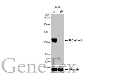
Wild-type (WT) and N-Cadherin knockout (KO) 293T cell extracts (30 μg) were separated by 5% SDS-PAGE, and the membrane was blotted with N-Cadherin antibody (GTX127345) diluted at 1:1000. The HRP-conjugated anti-rabbit IgG antibody (GTX213110-01) was used to detect the primary antibody.
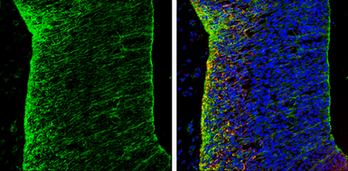
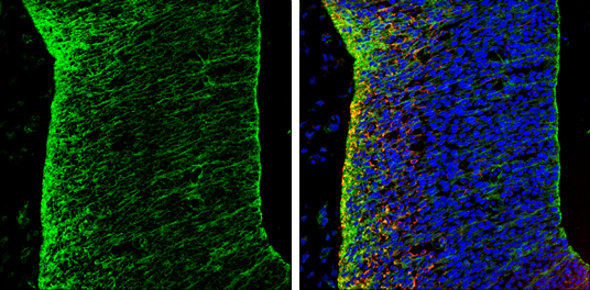
N-Cadherin antibody detects N-Cadherin protein expression by immunohistochemical analysis.
Sample: Frozen sectioned E13.5 Rat brain.
Green: N-Cadherin protein stained by N-Cadherin antibody (GTX127345) diluted at 1:250.
Red: beta Tubulin 3/ TUJ1, a mature neuron marker, stained by beta Tubulin 3/ TUJ1 antibody [GT11710] (GTX631836) diluted at 1:500.
Blue: Fluoroshield with DAPI (GTX30920).
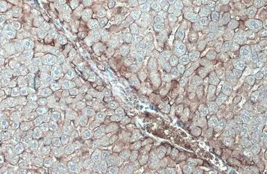
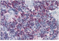
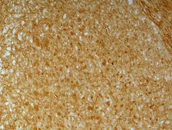
N-Cadherin antibody detects N-Cadherin protein at cell membrane by immunohistochemical analysis.
Sample: Paraffin-embedded mouse liver.
N-Cadherin stained by N-Cadherin antibody (GTX127345) diluted at 1:500.
Antigen Retrieval: Citrate buffer, pH 6.0, 15 min
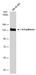
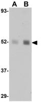
Whole cell extract (30 μg) was separated by 7.5% SDS-PAGE, and the membrane was blotted with N-Cadherin antibody (GTX127345) diluted at 1:1000. The HRP-conjugated anti-rabbit IgG antibody (GTX213110-01) was used to detect the primary antibody.
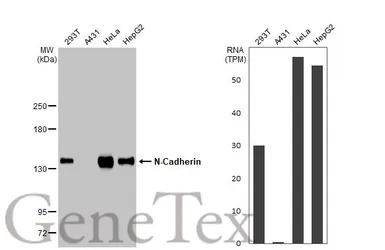
Various whole cell extracts (30 μg) were separated by 5% SDS-PAGE, and the membrane was blotted with N-Cadherin antibody (GTX127345) diluted at 1:1000. The HRP-conjugated anti-rabbit IgG antibody (GTX213110-01) was used to detect the primary antibody. Corresponding RNA expression data for the same cell lines are based on Human Protein Atlas program.
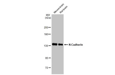
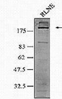
Various tissue extracts (50 μg) were separated by 5% SDS-PAGE, and the membrane was blotted with N-Cadherin antibody (GTX127345) diluted at 1:5000. The HRP-conjugated anti-rabbit IgG antibody (GTX213110-01) was used to detect the primary antibody.
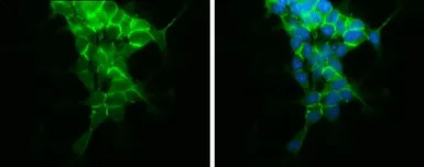
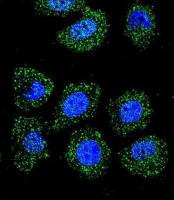
N-Cadherin antibody detects N-Cadherin protein at cell membrane by immunofluorescent analysis.
Sample: SH-SY5Y cells were fixed in 4% PFA at RT for 15 min.
Green: N-Cadherin protein stained by N-Cadherin antibody (GTX127345) diluted at 1:500.
Blue: Hoechst 33342 staining.
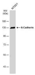
Whole cell extract (30 μg) was separated by 7.5% SDS-PAGE, and the membrane was blotted with N-Cadherin antibody (GTX127345) diluted at 1:1000. The HRP-conjugated anti-rabbit IgG antibody (GTX213110-01) was used to detect the primary antibody.
N-Cadherin antibody detects N-Cadherin protein on embryonic mouse brain by immunohistochemical analysis. Sample: Frozen section of embryonic mouse brain (mE18.5). N-Cadherin antibody (GTX127345) diluted at 1:500.
风险提示:丁香通仅作为第三方平台,为商家信息发布提供平台空间。用户咨询产品时请注意保护个人信息及财产安全,合理判断,谨慎选购商品,商家和用户对交易行为负责。对于医疗器械类产品,请先查证核实企业经营资质和医疗器械产品注册证情况。
 文献和实验
文献和实验Chen LH et al., BMC Cancer 2016 (PMID:27391030)
Kosim MY et al., Front Pharmacol 2022 (PMID:36199689)
Chua HH et al., Cell Mol Gastroenterol Hepatol 2023 (PMID:36191855)
Chiou HC et al., Antioxidants (Basel) 2023 (PMID:36830091)
Chang KS et al., Int J Mol Sci 2022 (PMID:36232736)
Lee WJ et al., J Food Drug Anal 2021 (PMID:35696218)
Wang L et al., Bioengineered 2022 (PMID:35609330)
Huang-Chi Chen et al., Front Pharmacol 2022 (PMID:35517780)
Mai Takahashi et al., Front Pharmacol 2021 (PMID:35058772)
Zhu X et al., Sci Adv 2022 (PMID:35245119)
Zou W et al., J Cell Mol Med 2022 (PMID:35229451)
Russell DF et al., Front Neuroanat 2020 (PMID:33192338)
Yang P et al., Exp Biol Med (Maywood) 2019 (PMID:31675905)
Lai YW et al., Cells 2021 (PMID:34440660)
Wang LT et al., Cancer Lett 2021 (PMID:34265398)
Belvedere R et al., Eur J Pharm Sci 2021 (PMID:34022411)
Milani M et al., J Neuroinflammation 2021 (PMID:34118929)
Nicolai A et al., Pharmaceuticals (Basel) 2021 (PMID:34204738)
Ou M et al., Oncol Rep 2021 (PMID:33955525)
Li Y et al., Transl Lung Cancer Res 2021 (PMID:34012797)
Milani M et al., Research Square 2021 (Epub)
Hu W et al., JCI Insight 2021 (PMID:33400686)
Chen YT et al., J Clin Invest 2021 (PMID:33465051)
Chen H.Y. et al., Cancers 2021;13(6)
Kuo TT et al., ACS Omega 2020 (PMID:33344833)
Duan C et al., Oncol Lett 2021 (PMID:33552262)
Kuo CH et al., Cell Biochem Funct 2020 (PMID:33135206)
Krzysiek-Maczka G et al., Microorganisms 2020 (PMID:33023180)
Ho KH et al., Cell Mol Neurobiol 2020 (PMID:33025417)
Wang LT et al., EMBO Rep 2020 (PMID:31908141)
Yang P et al., Int J Med Sci 2019 (PMID:31839751)
Marcinkiewcz CA et al., ACS Chem Neurosci 2019 (PMID:31140276)
Raineri A et al., Int J Mol Sci 2019 (PMID:31783660)
Lee JY et al., PLoS One 2020 (PMID:32730336)
Wang SC et al., Biochem Pharmacol 2020 (PMID:32679125)
Li Z et al., Front Cell Neurosci 2020 (PMID:32714149)
Le Saux G et al., ACS Appl Mater Interfaces 2020 (PMID:32323968)
Fang CH et al., J Biomed Sci 2020 (PMID:32169072)
Hsu SY et al., Onco Targets Ther 2019 (PMID:31571912)
Li-Chien Chang et al., Nutrition 2019 (Epub)
Kuo YL et al., Sci Rep 2019 (PMID:31311941)
Hung CY et al., Mol Neurobiol 2019 (PMID:31317491)
Lee DK et al., Biochem Biophys Res Commun 2019 (PMID:31076103)
Pei YF et al., Am J Pathol 2019 (PMID:30735628)
Mansel C et al., Neurotoxicol Teratol 2019 (PMID:30776472)
Chang CY et al., Cell Death Discov 2018 (PMID:30393570)
DaSilva-Arnold S et al., Mol Hum Reprod 2018 (PMID:30462321)
Chen CM et al., J Renin Angiotensin Aldosterone Syst 2018 (PMID:30264671)
Li JM et al., Ther Adv Med Oncol 2018 (PMID:30159048)
Hseu YC et al., J Cell Physiol 2018 (PMID:30146779)
Terasaki M et al., Anticancer Res 2018 (PMID:29599336)
Chen CY et al., Int J Mol Sci 2018 (PMID:29382035)
Shentu TP et al., Sci Rep 2017 (PMID:29273797)
Chen PC et al., J Cell Physiol 2017 (PMID:29215705)
Taglieri L et al., Invest New Drugs 2017 (PMID:28965307)
Chou HC et al., Toxicological Sciences 2017 (Epub)
Chan SM et al., Sci Rep 2017 (PMID:28819223)
Fujiki K et al., J Biol Chem 2017 (PMID:28302721)
Tong D et al., Cancer Lett 2016 (PMID:28043910)
Jian MY et al., Am J Physiol Lung Cell Mol Physiol 2016 (PMID:26545901)
Li CH et al., Arch Toxicol 2016 (PMID:27752740)
Jheng HF et al., Mol Nutr Food Res 2016 (PMID:27748993)
Application of Functional Blocking Antibodies: N-Cadherin and Chick Embryonic Limb Development
The current trend in developmental biology research is to identify candidate functional genes and then manipulate their expression or activity by either gain or loss of function to elucidate the specific roles of their protein products
Generation of Antibody Molecules Through Antibody Engineering
been overcome to a large extent using genetic-engineering techniques to produce chimeric mouse/human and completely human antibodies. Such an approach is particularly suitable because of the domain structure of the antibody molecule ( 2 ), where functional
The importance of antibody molecules was first recognized in the 1890s, when it was shown that immunity to tetanus and diphtheria was caused by antibodies against the bacterial exotoxins (1 ). Around the same time, it was shown that antisera
 技术资料
技术资料暂无技术资料 索取技术资料
