

- ¥1700 - 4000
- GeneTex

- 美国
- GTX104763
- 2025年07月16日
- WB, ICC/IF, IHC-P, IHC-Fr, FACS
- Rabbit
- Human

企业认证
相关产品推荐更多 >


![Estrogen Receptor alpha antibody [GT9004]](https://img1.dxycdn.com/2022/0328/907/3760111828182500453-14.jpg!wh200)
万千商家帮你免费找货
0 人在求购买到急需产品
- 详细信息
- 询价记录
- 文献和实验
- 技术资料
- 免疫原:
Carrier-protein conjugated synthetic peptide encompassing a sequence within the C-terminus region of human PD-L1. The exact sequence is proprietary.
- 亚型:
IgG
- 形态:
Liquid
- 保存条件:
Store as concentrated solution. Centrifuge briefly prior to opening vial. For short-term storage (1-2 weeks), store at 4ºC. For long-term storage, aliquot and store at -20ºC or below. Avoid multiple freeze-thaw cycles.
- 克隆性:
Polyclonal
- 标记物:
Unconjugated
- 适应物种:
Human
- 保质期:
12 months from the shipping date of the product.
- 抗原来源:
Human
- 目录编号:
GTX104763
- 级别:
Primary Antibodies
- 库存:
Available
- 供应商:
GeneTex
- 宿主:
Rabbit
- 应用范围:
WB, ICC/IF, IHC-P, IHC-Fr, FACS
- 浓度:
0.29 mg/ml (Please refer to the vial label for the specific concentration.)
- 靶点:
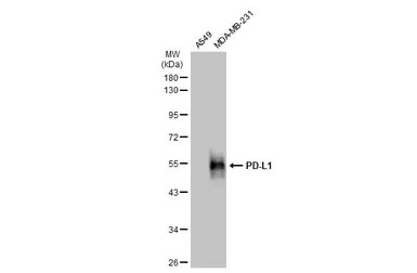
PD-L1
- 抗体英文名:
PD-L1 antibody
- 抗体名:
PD-L1 抗体
- 规格:
100 μl/25 μl
| 规格: | 100 μl | 产品价格: | ¥4000.0 |
|---|---|---|---|
| 规格: | 25 μl | 产品价格: | ¥1700.0 |
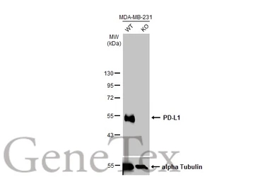
Wild-type (WT) and PD-L1 knockout (KO) MDA-MB-231 cell extracts (30 μg) were separated by 10% SDS-PAGE, and the membrane was blotted with PD-L1 antibody (GTX104763) diluted at 1:4000. The HRP-conjugated anti-rabbit IgG antibody (GTX213110-01) was used to detect the primary antibody.
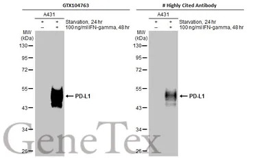
Untreated (–) and treated (+) A431 whole cell extracts (30 μg) were separated by 10% SDS-PAGE, and the membranes were blotted with PD-L1 antibody (GTX104763) diluted at 1:1200 and competitor's antibody (CST#13684) diluted at 1:500. The HRP-conjugated anti-rabbit IgG antibody (GTX213110-01) was used to detect the primary antibody.
*The competitor is not affiliated with GeneTex and does not endorse this product.
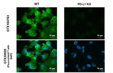
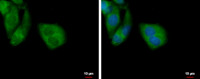
PD-L1 antibody detects PD-L1 protein at cell membrane by immunofluorescent analysis.
Sample: MDA-MB-231 cells were fixed in ice-cold MeOH for 5 min.
Green: PD-L1 stained by PD-L1 antibody (GTX104763) diluted at 1:500.
Blue: Fluoroshield with DAPI (GTX30920).
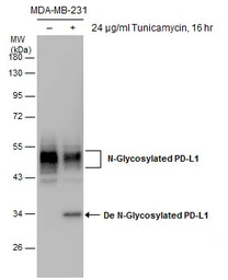
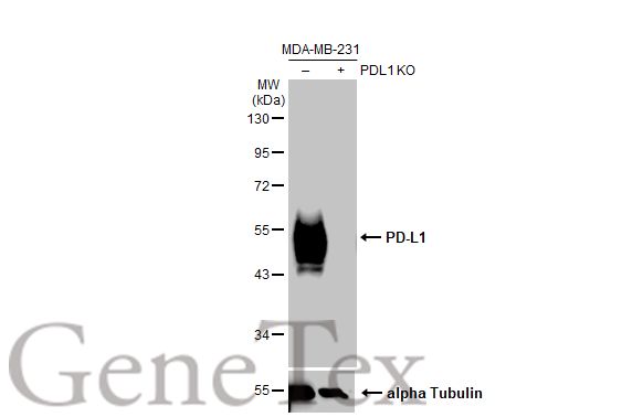
Untreated (–) and treated (+) MDA-MB-231 whole cell extracts (30 μg) were separated by 10% SDS-PAGE, and the membrane was blotted with PD-L1 antibody (GTX104763) diluted at 1:1000.
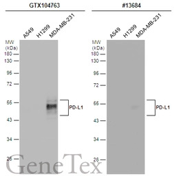
Various whole cell extracts (30 μg) were separated by 12% SDS-PAGE, and the membranes were blotted with PD-L1 antibody (GTX104763) diluted at 1:2000 and competitor's antibody (CST#13684) diluted by 1:500. The HRP-conjugated anti-rabbit IgG antibody (GTX213110-01) was used to detect the primary antibody.
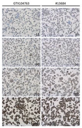
PD-L1 antibody detects PD-L1 protein at cell membrane in PD-L1 protein-expressing cell lines by immunohistochemical analysis. Antibodies: PD-L1 antibody (GTX104763) diluted at 1:1000, and competitor's antibody diluted at 1:50. Samples: Negative (-), low positive (+), intermediate positive (++) and strong positive (+++) cell line cores assessed using Quantitative Digital Pathology.
Antigen Retrieval: Citrate buffer, pH 6.0, 15 min
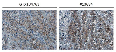
PD-L1 antibody detects PD-L1 protein at cell membrane in human ovarian carcinoma by immunohistochemical analysis. Antibodies: PD-L1 antibody (GTX104763) diluted at 1:1000, and competitor's antibody diluted at 1:50.
Antigen Retrieval: Citrate buffer, pH 6.0, 15 min
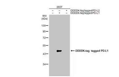
Non-transfected (–) and transfected (+) 293T whole cell extracts (30 μg) were separated by 10% SDS-PAGE, and the membrane was blotted with PD-L1 antibody (GTX104763) diluted at 1:1000. The HRP-conjugated anti-rabbit IgG antibody (GTX213110-01) was used to detect the primary antibody, and the signal was developed with Trident ECL plus-Enhanced.
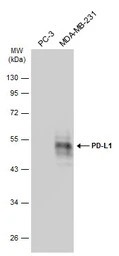
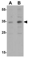
Various whole cell extracts (30 μg) were separated by 10% SDS-PAGE, and the membrane was blotted with PD-L1 antibody (GTX104763) diluted at 1:2000. The HRP-conjugated anti-rabbit IgG antibody (GTX213110-01) was used to detect the primary antibody, and the signal was developed with Trident ECL plus-Enhanced.
Various whole cell extracts (30 μg) were separated by 10% SDS-PAGE, and the membrane was blotted with PD-L1 antibody (GTX104763) diluted at 1:2000. The HRP-conjugated anti-rabbit IgG antibody (GTX213110-01) was used to detect the primary antibody.
风险提示:丁香通仅作为第三方平台,为商家信息发布提供平台空间。用户咨询产品时请注意保护个人信息及财产安全,合理判断,谨慎选购商品,商家和用户对交易行为负责。对于医疗器械类产品,请先查证核实企业经营资质和医疗器械产品注册证情况。
- 作者
- 内容
- 询问日期
 文献和实验
文献和实验Buisseret L et al., OncoImmunology 2016 (Epub)
Lin YM et al., PLoS One 2015 (PMID:26562534)
Wu Y et al., Nat Cancer 2023 (PMID:36894639)
Chung SY et al., Neoplasia 2023 (PMID:36442297)
Chung BS et al., Int J Mol Sci 2022 (PMID:36362062)
Su KW et al., Cells 2022 (PMID:36231010)
You-Zhe Lin et al., Comput Struct Biotechnol J 2022 (PMID:35024096)
Jesus Pacheco-Torres et al., Cancer Metab 2021 (PMID:33608051)
Hung YH et al., Am J Cancer Res 2022 (PMID:35261797)
Omenai SA et al., PLoS One 2022 (PMID:35139126)
Asrini R et al., Mol Clin Oncol 2022 (PMID:35003740)
Lin YZ et al., Computational and Structural Biotechnology Journal 2022 20()
Li L et al., Pharmacological Research - Modern Chinese Medicine 2021 1()
Tuminello S et al., Transl Lung Cancer Res 2020 (PMID:32953509)
Li L et al., Cancer Sci 2021 (PMID:33682294)
Qin G et al., Nat Commun 2020 (PMID:32245950)
Liu Z et al., Int J Clin Exp Pathol 2017 (PMID:31966537)
Nguyen HD et al., Cancers (Basel) 2019 (PMID:31835799)
Chou CW et al., Am J Cancer Res 2020 (PMID:32905506)
Huang TY et al., Cells 2020 (PMID:32756527)
Kim D et al., J Clin Med 2020 (PMID:32349330)
Lenouvel D et al., Oral Oncol 2020 (PMID:32330687)
Gondhowiardjo SA et al., PLoS One 2020 (PMID:32191754)
Pan MR et al., Cancers (Basel) 2019 (PMID:31905966)
Chan LC et al., J Clin Invest 2019 (PMID:31305264)
Polioudaki H et al., Cell Oncol (Dordr) 2019 (PMID:30680705)
Wei-faYang et al., Oral Oncology 2018 86()
Buisseret L et al., Oncoimmunology 2017 (PMID:28197375)
Cha JH et al., Mol Cell 2018 (PMID:30118680)
Li CW et al., Cancer Cell 2018 (PMID:29438695)
Takeuchi M et al., Immunol Lett 2018 (PMID:29366663)
Liu Z et al., lInt J Clin Exp Pathol 2017;10(12)
He B et al., Biomed Pharmacother 2017 (PMID:29247952)
Lin PL et al., Eur J Cancer 2017 (PMID:28892778)
Lin PL et al., Cancer Med 2017 (PMID:28795532)
Wang LT et al., Cancer Res 2017 (PMID:28625979)
Qing Y et al., Drug Des Devel Ther 2015 (PMID:25733810)
2015 年 12 月,美国前总统吉米 · 卡特在接受了靶向放疗和免疫抗癌新药 Keytruda(PD-1 抑制剂,MSD)治疗后,医生发现,在他的脑部磁共振扫描图像中,此前出现的黑色素瘤(Melanoma)与新生的癌细胞都彻底消失了。这种治疗效果引起了业内的广泛关注,也彰显了 PD-1/PD-L1 免疫疗法在肿瘤治疗中的巨大潜力。PD-1/PD-L1 是什么?1992 年,日本京都大学 Tasuku Honjo 教授首次报道并克隆了 PD-1 基因[1]。1999 年,华人学者陈列平教授报道
宽度:562 px高度:343 px维持原图长宽比:确认自 2011 年第一个免疫检验点 CTLA4 抑制剂伊匹单抗(Ipilimumab)获批上市以来,肿瘤免疫疗法越来越受到重视。特别是近几年陆续获批 PD1/PD-L1 单抗药物在临床治疗中的不断突破,肿瘤免疫治疗已从 1.0 时代进入 2.0 时代。免疫疗法适应症也在不断拓展,从黑色素瘤逐渐延伸到非小细胞肺癌、膀胱癌、头颈癌霍奇金淋巴瘤等癌症的治疗。针对 PD1/PD-L1 免疫检验点的单抗药物无疑是肿瘤免疫治疗中当之无愧的明星,其中 O
「神药」再显威!杨黄浩等让二甲双胍扮演 PD-L1 「单抗」角色,提高治疗效果
ICBs 的抗肿瘤效果。 该研究成果以 Reducing PD-L1 expression with a self-assembled nanodrug: an alternative to PD-L1 antibody for enhanced chemo-immunotherapy. 为题,发表在了 2021 年 1 月 1 日的 Theranostics 上 [1]。 图片来源:Theranostics 研究内容:此前有研究报道,二甲双胍可以抑制肿瘤进展,改善癌症患者的预后和生存率。此外,也有研
 技术资料
技术资料需要更多技术资料 索取更多技术资料

