


- ¥1700 - 4000
- GeneTex

- 美国
- GTX104557
- 2025年07月12日
- WB, ICC/IF, IHC-P, FACS, IP, ELISA
- Rabbit
- Human, Mouse, Rat

企业认证
相关产品推荐更多 >
![Dopamine Receptor D1 antibody [N1-2], N-term](https://img1.dxycdn.com/2022/0328/018/5777631261817200453-14.jpg!wh200)
![Transgelin antibody [GT336]](https://img1.dxycdn.com/2022/0329/016/2307622781765020453-14.jpg!wh200)
![HLA Class I antibody [MEM-147]](https://img1.dxycdn.com/2022/0328/517/8042020520740400453-14.jpg!wh200)
万千商家帮你免费找货
0 人在求购买到急需产品
- 详细信息
- 文献和实验
- 技术资料
- 免疫原:
Recombinant protein encompassing a sequence within the center region of human Glypican 1. The exact sequence is proprietary.
- 亚型:
IgG
- 形态:
Liquid
- 保存条件:
Store as concentrated solution. Centrifuge briefly prior to opening vial. For short-term storage (1-2 weeks), store at 4ºC. For long-term storage, aliquot and store at -20ºC or below. Avoid multiple freeze-thaw cycles.
- 克隆性:
Polyclonal
- 标记物:
Unconjugated
- 适应物种:
Human, Mouse, Rat
- 保质期:
12 months from the shipping date of the product.
- 抗原来源:
Human
- 目录编号:
GTX104557
- 级别:
Primary Antibodies
- 库存:
Available
- 供应商:
GeneTex
- 宿主:
Rabbit
- 应用范围:
WB, ICC/IF, IHC-P, FACS, IP, ELISA
- 浓度:
0.58 mg/ml (Please refer to the vial label for the specific concentration.)
- 靶点:
Glypican 1
- 抗体英文名:
Glypican 1 antibody [N3C3]
- 抗体名:
Glypican 1 抗体 [N3C3]
- 规格:
100 μl/25 μl
| 规格: | 100 μl | 产品价格: | ¥4000.0 |
|---|---|---|---|
| 规格: | 25 μl | 产品价格: | ¥1700.0 |
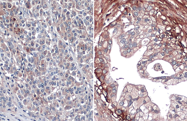
Glypican 1 antibody [N3C3] detects Glypican 1 protein at cell membrane by immunohistochemical analysis.
Sample: Paraffin-embedded human normal pancreas (left) and pancreatic cancer (right).
Glypican 1 stained by Glypican 1 antibody [N3C3] (GTX104557) diluted at 1:500.
Antigen Retrieval: Citrate buffer, pH 6.0, 15 min
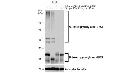
Untreated (–) and treated (+) A431 whole cell extracts (30 μg) were separated by 7.5% SDS-PAGE, and the membrane was blotted with Glypican 1 antibody [N3C3] (GTX104557) diluted at 1:3000. The HRP-conjugated anti-rabbit IgG antibody (GTX213110-01) was used to detect the primary antibody.
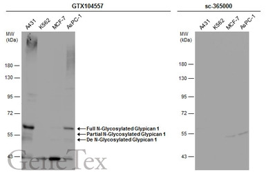
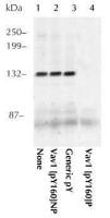
Various whole cell extracts (30 μg) were separated by 7.5% SDS-PAGE, and the membranes were blotted with Glypican 1 antibody [N3C3] (GTX104557) diluted at 1:500 and competitor's antibody (sc-365000) diluted at 1:500. The HRP-conjugated anti-rabbit IgG antibody (GTX213110-01) was used to detect the primary antibody.
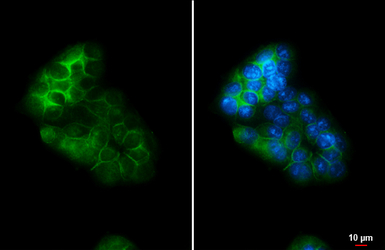
Glypican 1 antibody [N3C3] detects Glypican 1 protein at cell membrane and cytoplasm by immunofluorescent analysis.
Sample: MCF-7 cells were fixed in ice-cold MeOH for 5 min.
Green: Glypican 1 stained by Glypican 1 antibody [N3C3] (GTX104557) diluted at 1:500.
Blue: Hoechst 33342 staining.
Scale bar= 10 μm.
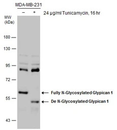
Untreated (–) and treated (+) MDA-MB-231 whole cell extracts (30 μg) were separated by 7.5% SDS-PAGE, and the membrane was blotted with Glypican 1 antibody [N3C3] (GTX104557) diluted at 1:1500. The HRP-conjugated anti-rabbit IgG antibody (GTX213110-01) was used to detect the primary antibody.
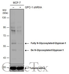
Non-transfected (–) and transfected (+) MCF-7 whole cell extracts (50 μg) were separated by 7.5% SDS-PAGE, and the membrane was blotted with Glypican 1 antibody [N3C3] (GTX104557) diluted at 1:1500. The HRP-conjugated anti-rabbit IgG antibody (GTX213110-01) was used to detect the primary antibody.
Untreated (–) and treated (+) A431 whole cell extracts (30 μg) were separated by 7.5% SDS-PAGE, and the membrane was blotted with Glypican 1 antibody [N3C3] (GTX104557) diluted at 1:500. The HRP-conjugated anti-rabbit IgG antibody (GTX213110-01) was used to detect the primary antibody.
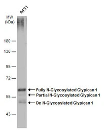
A431 whole cell extracts (30 μg) was separated by 7.5% SDS-PAGE, and the membrane was blotted with Glypican 1 antibody [N3C3] (GTX104557) diluted at 1:1500. The HRP-conjugated anti-rabbit IgG antibody (GTX213110-01) was used to detect the primary antibody.
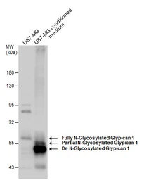
U87-MG whole cell extract and conditioned medium (30 μg) were separated by 7.5% SDS-PAGE, and the membrane was blotted with Glypican 1 antibody [N3C3] (GTX104557) diluted at 1:1500. The HRP-conjugated anti-rabbit IgG antibody (GTX213110-01) was used to detect the primary antibody.
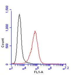
Glypican 1 antibody [N3C3] (GTX104557) detects Glypican 1 protein by flow cytometry analysis.
Sample: A431 cell.
Black: Unlabelled sample was used as a control.
Red: Glypican 1 antibody [N3C3] (GTX104557) dilution: 1:100.
Acquisition of 20,000 events were collected for FACS analysis.
风险提示:丁香通仅作为第三方平台,为商家信息发布提供平台空间。用户咨询产品时请注意保护个人信息及财产安全,合理判断,谨慎选购商品,商家和用户对交易行为负责。对于医疗器械类产品,请先查证核实企业经营资质和医疗器械产品注册证情况。
 文献和实验
文献和实验Huang G et al., Nat Commun 2017 (PMID:28102194)
Bologna-Molina R et al., Appl Immunohistochem Mol Morphol 2015 (PMID:25046223)
Sukhneeraj P Kaur et al., Sci Rep 2021 (PMID:33927256)
Munekage E et al., Neoplasia 2021 (PMID:34332450)
Zhang J et al., Ecotoxicol Environ Saf 2021 (PMID:33933809)
Emmanouilidi A et al., Proteomics 2019 (PMID:30893511)
P Kim et al., Transl Psychiatry 2019 (PMID:30664618)
Lu H et al., Cancer Med 2017 (PMID:28440066)
Qian JY et al., Oncology Letters 2018 (Epub)
Hsia K et al., Acta Biomater 2017 (PMID:28110073)
In the field of therapeutic recombinant proteins, monoclonal antibodies (mAbs) have achieved a rising success with more than 30 mAbs that have reached the market in the past 20 years. From a structural standpoint, one of the most important
Materials 0.1M NaHC03 pH9 DMSO NHS-Biotin (N-Hydroxysuccinimidobiotin, Sigma #H-1759) PBS Procedure Dialyze the sample against carbonate buffer. After dialysis, adjust
Monoclonal Antibody Production Protocol
the rinse twice. Add 100 ul of blocking solution to every well, leave 1 hr at room Temp or O.N at 4°C. PRIMARY ANTIBODY Add the antibody to be tested: Sup of cells = 25 ul, mix well by pipetting up and down (10 times).serum, ascites = 1:100 and a series
 技术资料
技术资料暂无技术资料 索取技术资料

