

- ¥1700 - 4000
- GeneTex

- 美国
- GTX102640
- 2025年07月10日
- WB, ICC/IF, IHC-P, IP
- Rabbit
- Human, Mouse, Rat

企业认证
相关产品推荐更多 >
![Cyclophilin E antibody [AT17E8]](https://img1.dxycdn.com/2022/0328/192/3430167378450800453-14.jpg!wh200)
![CXCL8 / IL8 antibody [3E4D8]](https://img1.dxycdn.com/2022/0328/441/4396843623877500453-14.jpg!wh200)
万千商家帮你免费找货
0 人在求购买到急需产品
- 详细信息
- 文献和实验
- 技术资料
- 免疫原:
Recombinant protein encompassing a sequence within the C-terminus region of human Topoisomerase II beta. The exact sequence is proprietary.
- 亚型:
IgG
- 形态:
Liquid
- 保存条件:
Store as concentrated solution. Centrifuge briefly prior to opening vial. For short-term storage (1-2 weeks), store at 4ºC. For long-term storage, aliquot and store at -20ºC or below. Avoid multiple freeze-thaw cycles.
- 克隆性:
Polyclonal
- 标记物:
Unconjugated
- 适应物种:
Human, Mouse, Rat
- 保质期:
12 months from the shipping date of the product.
- 抗原来源:
Human
- 目录编号:
GTX102640
- 级别:
Primary Antibodies
- 库存:
Available
- 供应商:
GeneTex
- 宿主:
Rabbit
- 应用范围:
WB, ICC/IF, IHC-P, IP
- 浓度:
0.48 mg/ml (Please refer to the vial label for the specific concentration.)
- 靶点:
Topoisomerase II beta
- 抗体英文名:
Topoisomerase II beta antibody [C3], C-term
- 抗体名:
Topoisomerase II beta 抗体 [C3], C-term
- 规格:
100 μl/25 μl
| 规格: | 100 μl | 产品价格: | ¥4000.0 |
|---|---|---|---|
| 规格: | 25 μl | 产品价格: | ¥1700.0 |
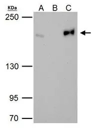
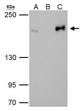
Topoisomerase II beta antibody immunoprecipitates Topoisomerase II beta protein in IP experiments. IP Sample: HeLa whole cell lysate/extract A. 40 μg HeLa whole cell lysate/extract B. Control with 2 μg of preimmune rabbit IgG C. Immunoprecipitation of Topoisomerase II beta protein by 2 μg of Topoisomerase II beta antibody (GTX102640) 5% SDS-PAGE The immunoprecipitated Topoisomerase II beta protein was detected by Topoisomerase II beta antibody (GTX102640) diluted at 1:1000. EasyBlot anti-rabbit IgG (GTX221666-01) was used as a secondary reagent.
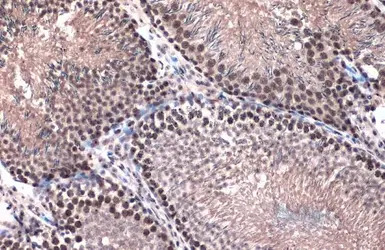
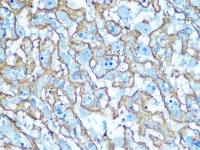
Topoisomerase II beta antibody [C3], C-term detects Topoisomerase II beta protein at nucleus by immunohistochemical analysis.
Sample: Paraffin-embedded rat testis.
Topoisomerase II beta stained by Topoisomerase II beta antibody [C3], C-term (GTX102640) diluted at 1:500.
Antigen Retrieval: Citrate buffer, pH 6.0, 15 min
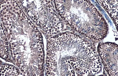
Topoisomerase II beta antibody [C3], C-term detects Topoisomerase II beta protein at nucleus by immunohistochemical analysis.
Sample: Paraffin-embedded mouse testis.
Topoisomerase II beta stained by Topoisomerase II beta antibody [C3], C-term (GTX102640) diluted at 1:500.
Antigen Retrieval: Citrate buffer, pH 6.0, 15 min
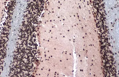
Topoisomerase II beta antibody [C3], C-term detects Topoisomerase II beta protein at nucleus by immunohistochemical analysis.
Sample: Paraffin-embedded mouse brain.
Topoisomerase II beta stained by Topoisomerase II beta antibody [C3], C-term (GTX102640) diluted at 1:500.
Antigen Retrieval: Citrate buffer, pH 6.0, 15 min

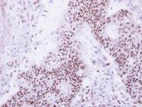
Immunohistochemical analysis of paraffin-embedded human ovarian cancer, using Topoisomerase II beta(GTX102640) antibody at 1:250 dilution.
Antigen Retrieval: Trilogy™ (EDTA based, pH 8.0) buffer, 15min
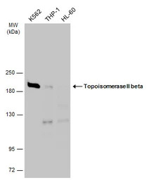
Various whole cell extracts (30 μg) were separated by 5% SDS-PAGE, and the membrane was blotted with Topoisomerase II beta antibody [C3], C-term (GTX102640) diluted at 1:3000. The HRP-conjugated anti-rabbit IgG antibody (GTX213110-01) was used to detect the primary antibody.
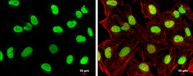
Topoisomerase II beta antibody [C3], C-term detects Topoisomerase II beta protein at nucleus by immunofluorescent analysis.
Sample: HeLa cells were fixed in 4% PFA at RT for 15 min.
Green: Topoisomerase II beta stained by Topoisomerase II beta antibody [C3], C-term (GTX102640) diluted at 1:500.
Red: phalloidin, a cytoskeleton marker, diluted at 1:100.
Scale bar= 10 μm.
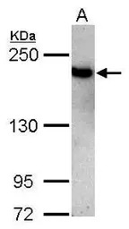
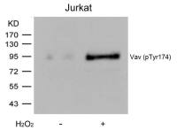
Sample (30 μg of whole cell lysate)
A: Jurkat
5% SDS PAGE
GTX102640 diluted at 1:3000
The HRP-conjugated anti-rabbit IgG antibody (GTX213110-01) was used to detect the primary antibody.
风险提示:丁香通仅作为第三方平台,为商家信息发布提供平台空间。用户咨询产品时请注意保护个人信息及财产安全,合理判断,谨慎选购商品,商家和用户对交易行为负责。对于医疗器械类产品,请先查证核实企业经营资质和医疗器械产品注册证情况。
 文献和实验
文献和实验Chen JC et al., Biomolecules 2019 (PMID:31505885)
Liu TP et al., R Soc Open Sci 2018 (PMID:30564416)
Hangas A et al., Nucleic Acids Res 2018 (PMID:30169847)
secondary antibody review -- data from 99 publications
cytometry used as a control to detect cell responses targeted antigen 7 Alexa Fluor 488 7 Cy3 8 goat IgG Alexa Fluor 488 1:2000 detect antibody binding in human embryonic kidney 293T cells Invitrogen 9 donkey
1E C-term Antibody AP8428a PTPIA2 N-term Antibody AP8437a DUSP6 N-term Antibody AP8457a PPM1E N-term Antibody AP8427a PTPIA2beta N-term Antibody AP8438b DUSP7 C-term Antibody AP8447b PPM1F C-term Antibody AP8417a PTPkappa N-term Antibody AP
祝贺〖雅裕睿安〗成为Signalway Antibody(SAB)独家
ul; 货号21617,产品描述:Tubulin beta-III Antibody;规格 50 ul / 100 ul; 货号21612,产品描述:GAPDH Antibody;规格 50 ul / 100 ul; 货号21335,产品描述:β-Tubulin mouse mAb;规格 50 ul / 100 ul; 货号21336,产品描述:GAPDH mouse
 技术资料
技术资料暂无技术资料 索取技术资料

