


- ¥1700 - 4000
- GeneTex

- 美国
- GTX101507
- 2025年07月06日
- WB, ICC/IF, IHC-P, IHC-Fr, FACS, IP
- Rabbit
- Human, Mouse, Rat, Frog, Ceratotherium simum

企业认证

研选同类产品更多 >
![Enterovirus D68 VP1 antibody [HL1997]](https://img1.dxycdn.com/2023/0605/423/2302796740531755661-14.jpg!wh200)
![p73 antibody [5B429]](https://img1.dxycdn.com/2022/0328/857/9527819040816400453-14.jpg!wh200)
万千商家帮你免费找货
0 人在求购买到急需产品
- 详细信息
- 用户评价
- 文献和实验
- 技术资料
- 免疫原:
Recombinant protein encompassing a sequence within the center region of human SOX2. The exact sequence is proprietary.
- 亚型:
IgG
- 形态:
Liquid
- 保存条件:
Store as concentrated solution. Centrifuge briefly prior to opening vial. For short-term storage (1-2 weeks), store at 4ºC. For long-term storage, aliquot and store at -20ºC or below. Avoid multiple freeze-thaw cycles.
- 克隆性:
Polyclonal
- 标记物:
Unconjugated
- 适应物种:
Human, Mouse, Rat, Frog, Ceratotherium simum
- 保质期:
12 months from the shipping date of the product.
- 抗原来源:
Human
- 目录编号:
GTX101507
- 级别:
Primary Antibodies
- 库存:
Available
- 供应商:
GeneTex
- 宿主:
Rabbit
- 应用范围:
WB, ICC/IF, IHC-P, IHC-Fr, FACS, IP
- 浓度:
0.15 mg/ml (Please refer to the vial label for the specific concentration.)
- 靶点:
SOX2
- 抗体英文名:
SOX2 antibody [N1C3]
- 抗体名:
SOX2 抗体 [N1C3]
- 规格:
100 μl/25 μl
| 规格: | 100 μl | 产品价格: | ¥4000.0 |
|---|---|---|---|
| 规格: | 25 μl | 产品价格: | ¥1700.0 |
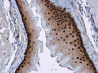
SOX2 antibody [N1C3] detects SOX2 protein at nucleus in mouse esophagus by immunohistochemical analysis.
Sample: Paraffin-embedded mouse esophagus.
SOX2 antibody [N1C3] (GTX101507) diluted at 1:500.
Antigen Retrieval: Citrate buffer, pH 6.0, 15 min

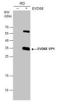
Whole cell extract (30 μg) was separated by 12% SDS-PAGE, and the membranes were blotted with SOX2 antibody [N1C3] (GTX101507) diluted at 1:1000 and competitor's antibody (sc-17320) diluted at 1:500. The HRP-conjugated anti-rabbit IgG antibody (GTX213110-01) was used to detect the primary antibody.
*The competitor is not affiliated with GeneTex and does not endorse this product.
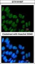
Immunofluorescence analysis of PFA-fixed Human ESC, using SOX2(GTX101507) antibody at 1:160 dilution.
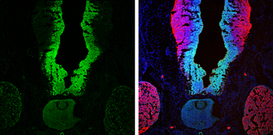
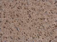
SOX2 antibody [N1C3] detects SOX2 protein at nucleus by immunohistochemical analysis.
Sample: Frozen sectioned E13.5 rat brain.
Green: SOX2 protein stained by SOX2 antibody [N1C3] (GTX101507) diluted at 1:250.
Red: beta Tubulin 3/ TUJ1, a mature neuron marker, stained by beta Tubulin 3/ TUJ1 antibody [GT11710] (GTX631836) diluted at 1:250.
Blue: Fluoroshield with DAPI (GTX30920).
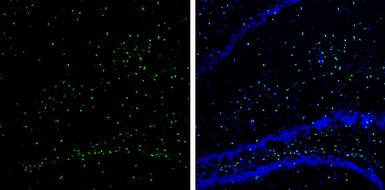
SOX2 antibody [N1C3] detects SOX2 protein expression by immunohistochemical analysis.
Sample: Frozen-sectioned adult mouse hippocampus.
Green: SOX2 protein stained by SOX2 antibody [N1C3] (GTX101507) diluted at 1:250.
Blue: Fluoroshield with DAPI (GTX30920).
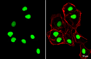
SOX2 antibody [N1C3] detects SOX2 protein at nucleus by immunofluorescent analysis.
Sample: NT2D1 cells were fixed in 4% PFA at RT for 15 min.
Green: SOX2 stained by SOX2 antibody [N1C3] (GTX101507) diluted at 1:500.
Red: phalloidin, a cytoskeleton marker, diluted at 1:100.
Scale bar= 10 μm.
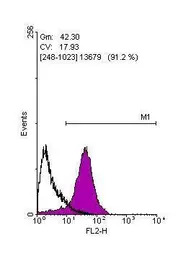
Flow cytometry on human embryonic stem cells, staining with SOX2 (GTX101507)antibody at 1:100 dilution(purple) or rabbit IgG (black).

Immunohistochemical analysis of paraffin-embedded Cal27 xenograft, using SOX2(GTX101507) antibody at 1:100 dilution.
Antigen Retrieval: Trilogy™ (EDTA based, pH 8.0) buffer, 15min
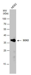
SOX2 antibody detects SOX2 protein by Western blot analysis. Whole cell extracts (30 μg) was separated by 12% SDS-PAGE, and the membrane was blotted with SOX2 antibody (GTX101507) at a dilution of 1:2500.

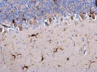
SOX2 antibody [N1C3] detects SOX2 protein at nucleus on rat brain stem by immunohistochemical analysis.
Sample: Paraffin-embedded rat brain stem.
SOX2 antibody [N1C3] (GTX101507) dilution: 1:500.
Antigen Retrieval: Trilogy™ (EDTA based, pH 8.0) buffer, 15min
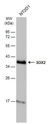
Whole cell extract (30 μg) was separated by 12% SDS-PAGE, and the membrane was blotted with SOX2 antibody [N1C3] (GTX101507) diluted at 1:10000. The HRP-conjugated anti-rabbit IgG antibody (GTX213110-01) was used to detect the primary antibody.
风险提示:丁香通仅作为第三方平台,为商家信息发布提供平台空间。用户咨询产品时请注意保护个人信息及财产安全,合理判断,谨慎选购商品,商家和用户对交易行为负责。对于医疗器械类产品,请先查证核实企业经营资质和医疗器械产品注册证情况。
 用户评价
用户评价 暂无用户评价
 文献和实验
文献和实验Misuno K et al., Stem Cell Res Ther 2013 (PMID:24423398)
Chen A et al., Front Mol Neurosci 2023 (PMID:37089691)
Jin C et al., Bioeng Transl Med 2023 (PMID:36925694)
Mohankumar K et al., Mol Carcinog 2022 (PMID:34699643)
Masola V et al., Front Oncol 2022 (PMID:35965510)
Peng Ye et al., Biomolecules 2022 (PMID:35625596)
Elisa De Tomi et al., Int J Mol Sci 2022 (PMID:35163570)
Wei-Fang Chang et al., Int J Mol Sci 2021 (PMID:33440839)
Cristina Mir et al., Cancers (Basel) 2021 (PMID:34638436)
Gupta A et al., Life (Basel) 2021 (PMID:34947952)
Ting HC et al., Cells 2021 (PMID:34685754)
Wang SM et al., Cell Death Discov 2021 (PMID:33436575)
Tsai LH et al., Sci Rep 2020 (PMID:33082357)
Chu EP et al., Stem Cell Res 2020 (PMID:33096384)
Carelli S et al., Nanotheranostics 2021 (PMID:33391972)
Hsu HS et al., J Biomed Sci 2020 (PMID:31928533)
Chen YL et al., Cancers (Basel) 2019 (PMID:31878324)
Blanas A et al., Cancers (Basel) 2020 (PMID:32927726)
Zhang G et al., Cancer Sci 2020 (PMID:32885530)
Yang SC et al., FASEB J 2019 (PMID:31242772)
Chi HC et al., Oncogene 2020 (PMID:32753649)
Wang J et al., Front Cell Neurosci 2020 (PMID:33192328)
Kerschner JL et al., J Cell Mol Med 2020 (PMID:32692488)
Chiou HC et al., Neoplasia 2020 (PMID:32438306)
Wang IH et al., Cancers (Basel) 2020 (PMID:32349352)
He X et al., Stem Cell Res Ther 2020 (PMID:32299508)
Zhao T et al., Stem Cell Reports 2020 (PMID:32160522)
Wang CH et al., Sci Rep 2020 (PMID:32042022)
Li X et al., J Cancer 2020 (PMID:32201526)
Fletcher PA et al., Front Endocrinol (Lausanne) 2019 (PMID:31620083)
Chang WF et al., Int J Mol Sci 2019 (PMID:30870992)
Chien CH et al., J Biomed Sci 2019 (PMID:31629402)
Bayat Mokhtari R et al., BMC Cancer 2019 (PMID:31470802)
Chen YH et al., Sci Rep 2019 (PMID:31289326)
Wang L et al., Toxicol Appl Pharmacol 2019 (PMID:31108107)
Yang Y et al., Neurochem Res 2019 (PMID:30628018)
Mansel C et al., Neurotoxicol Teratol 2019 (PMID:30776472)
Zhong PQ et al., Cell Signal 2019 (PMID:30448346)
Shen Z et al., Elife 2017 (PMID:28537559)
Ye Y et al., Neural Plast 2016 (PMID:28018679)
Hsu CC et al., J Biomed Sci 2016 (PMID:27863490)
Hildebrandt TB et al., Nat Commun 2018 (PMID:29973581)
Fawal MA et al., Cell Rep 2018 (PMID:29874574)
Yoshimura A et al., Dis Model Mech 2017 (PMID:29208635)
Wang AM et al., Oncotarget 2014 (PMID:24970812)
Ko CY et al., Mol Neurobiol 2017 (PMID:28478507)
Lin HC et al., J Agric Food Chem 2017 (PMID:27997180)
Gong W et al., Am J Cancer Res 2017 (PMID:28401007)
Chen MW et al., Cancer Res 2017 (PMID:28209618)
Gazarian KG et al., PLoS One 2017 (PMID:28125654)
Zhao T et al., PLoS One 2015 (PMID:26372456)
Mineda K et al., Stem Cells Transl Med 2015 (PMID:26494781)
Chen H et al., Proc Natl Acad Sci U S A 2015 (PMID:26483458)
Ye Y et al., Neural Plasticity 2016 (Epub)
Chang WF et al., PLoS One 2016 (PMID:27802323)
Chen MJ et al., Sci Rep 2016 (PMID:27713506)
Petrova A et al., Stem Cells Dev 2016 (PMID:27460132)
Zhang W et al., Sci Rep 2016 (PMID:27226076)
Chen CH et al., Anim Biotechnol 2016 (PMID:26980563)
Hung YH et al., Mol Neurobiol 2014 (PMID:25502463)
Emhemmed F et al., Biochem Pharmacol 2014 (PMID:24607276)
In the field of therapeutic recombinant proteins, monoclonal antibodies (mAbs) have achieved a rising success with more than 30 mAbs that have reached the market in the past 20 years. From a structural standpoint, one of the most important
Materials 0.1M NaHC03 pH9 DMSO NHS-Biotin (N-Hydroxysuccinimidobiotin, Sigma #H-1759) PBS Procedure Dialyze the sample against carbonate buffer. After dialysis, adjust
Monoclonal Antibody Production Protocol
the rinse twice. Add 100 ul of blocking solution to every well, leave 1 hr at room Temp or O.N at 4°C. PRIMARY ANTIBODY Add the antibody to be tested: Sup of cells = 25 ul, mix well by pipetting up and down (10 times).serum, ascites = 1:100 and a series
 技术资料
技术资料暂无技术资料 索取技术资料

