

- ¥1700 - 4000
- GeneTex

- 美国
- GTX100065
- 2025年07月16日
- WB, ICC/IF, IHC-P
- Rabbit
- Human, Mouse

企业认证
相关产品推荐更多 >
![eIF2 alpha antibody [2E4]](https://img1.dxycdn.com/2022/0328/992/0830010718232700453-14.jpg!wh200)

![GOT1 antibody [GT638]](https://img1.dxycdn.com/2022/0329/432/8759483482216020453-14.jpg!wh200)
万千商家帮你免费找货
0 人在求购买到急需产品
- 详细信息
- 文献和实验
- 技术资料
- 免疫原:
Carrier-protein conjugated synthetic peptide surrounding phospho Ser345 of human Chk1. The exact sequence is proprietary.
- 亚型:
IgG
- 形态:
Liquid
- 保存条件:
Store as concentrated solution. Centrifuge briefly prior to opening vial. For short-term storage (1-2 weeks), store at 4ºC. For long-term storage, aliquot and store at -20ºC or below. Avoid multiple freeze-thaw cycles.
- 克隆性:
Polyclonal
- 标记物:
Unconjugated
- 适应物种:
Human, Mouse
- 保质期:
12 months from the shipping date of the product.
- 抗原来源:
Human
- 目录编号:
GTX100065
- 级别:
Primary Antibodies
- 库存:
Available
- 供应商:
GeneTex
- 宿主:
Rabbit
- 应用范围:
WB, ICC/IF, IHC-P
- 浓度:
1.66 mg/ml (Please refer to the vial label for the specific concentration.)
- 靶点:
Chk1 (phospho Ser345)
- 抗体英文名:
Chk1 (phospho Ser345) antibody [C1C2], Internal
- 抗体名:
Chk1 (phospho Ser345) 抗体 [C1C2], Internal
- 规格:
100 μl/25 μl
| 规格: | 100 μl | 产品价格: | ¥4000.0 |
|---|---|---|---|
| 规格: | 25 μl | 产品价格: | ¥1700.0 |
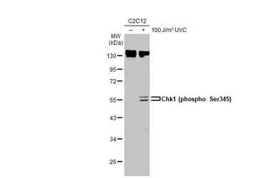
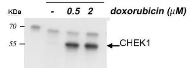
Untreated (–) and treated (+) C2C12 whole cell extract (30 μg) were separated by 10% SDS-PAGE, and the membrane was blotted with Chk1 (phospho Ser345) antibody [C1C2], Internal (GTX100065) diluted at 1:1000. The HRP-conjugated anti-rabbit IgG antibody (GTX213110-01) was used to detect the primary antibody, and the signal was developed with Trident ECL plus-Enhanced.
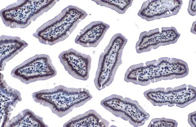
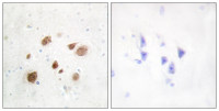
Chk1 (phospho Ser345) antibody [C1C2], Internal detects Chk1 (phospho Ser345) protein at nucleus by immunohistochemical analysis.
Sample: Paraffin-embedded mouse intestine.
Chk1 (phospho Ser345) stained by Chk1 (phospho Ser345) antibody [C1C2], Internal (GTX100065) diluted at 1:500.
Antigen Retrieval: Citrate buffer, pH 6.0, 15 min
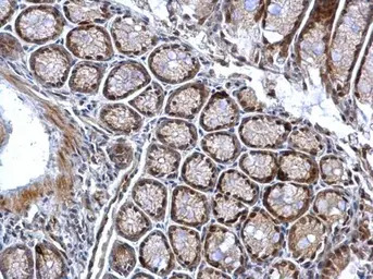
Chk1 (phospho Ser345) antibody [C1C2], Internal detects Chk1 (phospho Ser345) protein at nucleus on mouse colon by immunohistochemical analysis.
Sample: Paraffin-embedded mouse colon.
Chk1 (phospho Ser345) antibody [C1C2], Internal (GTX100065) dilution: 1:500.
Antigen Retrieval: Trilogy™ (EDTA based, pH 8.0) buffer, 15min
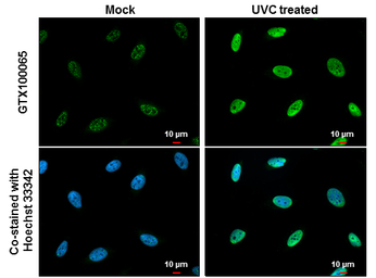
Chk1 (phospho Ser345) antibody [C1C2], Internal detects Chk1 (phospho Ser345) protein at nucleus by immunofluorescent analysis.
Samples: HeLa cells mock (left) and treated with 100 J/m2 UVC and recover for 8 hrs (right) were fixed in 4% PFA at RT for 15 min.
Green: Chk1 (phospho Ser345) protein stained by Chk1 (phospho Ser345) antibody [C1C2], Internal (GTX100065) diluted at 1:500.
Blue: Hoechst 33342 staining.
Scale bar = 10 μm.
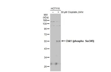
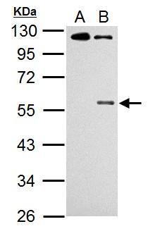
Untreated (–) and treated (+) HCT116 whole cell extracts (30 μg) were separated by 10% SDS-PAGE, and the membrane was blotted with Chk1 (phospho Ser345) antibody [C1C2], Internal (GTX100065) diluted at 1:500. The HRP-conjugated anti-rabbit IgG antibody (GTX213110-01) was used to detect the primary antibody, and the signal was developed with Trident ECL plus-Enhanced.
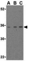
Sample: 20 μg of HCT116 P53 whole cell lysate 10% SDS PAGE
Chk1-phopho-S345 antibody
GTX100065 diluted at 1:1000
The HRP-conjugated anti-rabbit IgG antibody (GTX213110-01) was used to detect the primary antibody.

Immunohistochemical analysis of paraffin-embedded HeLa xenograft, using Chk1 (phospho Ser345) (GTX100065) antibody at 1:500 dilution.
Antigen Retrieval: Trilogy™ (EDTA based, pH 8.0) buffer, 15min
风险提示:丁香通仅作为第三方平台,为商家信息发布提供平台空间。用户咨询产品时请注意保护个人信息及财产安全,合理判断,谨慎选购商品,商家和用户对交易行为负责。对于医疗器械类产品,请先查证核实企业经营资质和医疗器械产品注册证情况。
 文献和实验
文献和实验Subramanian GN et al., J Cell Biol 2020 (PMID:32328643)
Luo J et al., mBio 2020 (PMID:32071277)
Hung CC et al., Oncotarget 2015 (PMID:25686830)
Yang C et al., Oncol Lett 2022 (PMID:35317026)
Sakata R et al., Cell Rep 2021 (PMID:33909997)
Methods for Studying Checkpoint Kinases Chk1
-terminal regulatory domain. Within the regulatory domain there are two residues, Serine-317 (S317) and Serine-345 (S345), which are phosphorylated in active Chk1 molecules, and subsequently dephosphorylated to inactivate Chk1 and allow mitotic entry
Mammalian CHK1 is a Ser/Thr effector kinase that plays critical roles in the DNA damage-activated cell cycle checkpoint signaling pathway downstream of ATR (ATM and Rad3-related protein kinase). This chapter is focused on describing an assay
Using Phospho‐Motif Antibodies to Determine Kinase Substrates
comprising both the phosphorylated residue and the surrounding residues that determine kinase specificity, with degenerate residues taking up the remaining positions. Currently, several categories of phospho?motif antibody are commercially available
 技术资料
技术资料暂无技术资料 索取技术资料

