


- ¥3975
- Agrisera
- 货号AS05 084
- 美国
- 2026年05月27日
- western blot (WB)
- 兔
- 植物

企业认证
相关产品推荐更多 >


![HA tag Antibody (109B2)[Biotin] , mAb, Rabbit,兔单抗(生物素标记 HA 标签抗体 109B2)](https://img1.dxycdn.com/p/s14/2026/0615/544/4285636518199236602.png!wh200)


万千商家帮你免费找货
0 人在求购买到急需产品
- 详细信息
- 文献和实验
- 技术资料
- 抗体名:
兔抗光系统II 核心蛋白D1多克隆抗体
- 抗体英文名:
PsbA|D1 protein of PSII,C-terminal
- 浓度:
见说明书
- 应用范围:
western blot (WB)
- 宿主:
兔
- 适应物种:
植物
- 标记物:
无
- 克隆性:
多抗/单行
- 保存条件:
低温
- 亚型:
IgG
- 规格:
100 µl
供应商:上海经科化学科技有限公司
服务热线:400-0199-638
QQ:472482400(上海经科)
微信号:shjkchem
活动:消费积分可换充值卡!
兔抗光系统II 核心蛋白D1多克隆抗体介绍:
货 号:AS05 084
中文名称:兔抗光系统II 核心蛋白D1多克隆抗体
英文名称:PsbA|D1 protein of PSII,C-terminal
应用:western blot (WB)
规格:100 µl
价格:3975元
兔抗光系统II 核心蛋白D1多克隆抗体简介:
|
|||||||||||||||||||||||||||||||||||||||||
| APPLICATION INFORMATION | ||
| recommended dilution | 1: 10 000 with standard ECL (WB) |
|
| expected | apparent MW | 38 | 28-30 kDa |
|
| confirmed reactivity | Anabaena 7120, Arabidopsis thaliana, Artemisia annua, Arundo sp., Chlamydomonas reinhardtii, Chromera velia, Colobanthus quitensis Kunt Bartl, Coscinodiscus wailesii, Craterostigma sp., Ditylum brightwellii, Glycine max, Hordeum vulgare, Lindernia sp., Marchantia polymorpha (liverwort),Miscanthus x giganteus, Microcystis aeruginosa, Nicotiana benthamiana, Panicum miliaceum, Panax ginseng, Panicum maximum, Paulinella chromatophora (amoeba), Physcomitrella patens, Pinus strobus, Prochlorococcus sp. (surface and deep water ecotype), Spartina alterniflora, Spirodela polyrhiza, Symbiodinium sp, Synechococcus sp. PCC 7942, Triticum aestivum, Zea mays |
|
| predicted reactivity | Lycopersicum esculentum, Medicago Sativa, Pisum Sativum and other di and monocots,conifers, brown and red algae, cyanobacteria; cellular [compartment marker] of thylakoid membrane |
|
| not reactive in | no confirmed exceptions from predicted reactivity known in the moment |
|
| additional information | The antibody is appropriate for detecting both, 24 kDa or the 10 kDa C-terminal fragments, whichever is generated under given treatment conditions. In our analysis we have seen both, ca. 24 kDa and ca. 10 kDa fragments from different samples, depending on treatments and isolation procedures. Rabbit anti-PsbA antibody can detect more than one band of PsbA protein, e.g. precursor and mature protein as compare to the hen anti-PsbA antibodies AS01 016. This antibody will detect the phosphorylated form of D1 as an alternate band to the main band on a high resolution gel. |
|
| selected references | Heinnickel et al. (2016). Tetratricopeptide repeat protein protects photosystem I from oxidative disruption during assembly. Proc Natl Acad Sci U S A. 2016 Mar 8;113(10):2774-9. doi: 10.1073/pnas.1524040113. Treves et al. (2016). The mechanisms whereby the green alga Chlorella ohadii, isolated from desert soil crust, exhibits unparalleled photodamage resistance. New Phytol. 2016 Feb 8. doi: 10.1111/nph.13870 Wang et al. (2015). The combined effects of UV-C radiation and H2O2 on Microcystis aeruginosa, a bloom-forming cyanobacterium. Chemosphere. 2015 Jun 16;141:34-43. doi: 10.1016/j.chemosphere.2015.06.020. Bancel et al. (2015). Proteomic Approach to Identify Nuclear Proteins in Wheat Grain. J Proteome Res. 2015 Sep 8. Vandenhecke et al. (2015). Changes in the Rubisco to photosystem ratio dominates photoacclimation across phytoplankton taxa. Photosynth Res. 2015 Jun;124(3):275-91. doi: 10.1007/s11120-015-0137-6. Epub 2015 Apr 11. Charuvi et al. (2015). Photoprotection Conferred by Changes in Photosynthetic Protein Levels and Organization during Dehydration of a Homoiochlorophyllous Resurrection Plant. Plant Physiol. 2015 Apr;167(4):1554-65. doi: 10.1104/pp.114.255794. Spence et al. (2014). Transcriptional responses indicate maintenance of photosynthetic proteins as key to the exceptional chilling tolerance of C4 photosynthesis in Miscanthus × giganteus. J Exp Bot. 2014 Jul;65(13):3737-47. doi: 10.1093/jxb/eru209. Epub 2014 Jun 22. Pandey and Pandey-Rai (2014). Modulations of physiological responses and possible involvement of defense-related secondary metabolites in acclimation of Artemisia annua L. against short-term UV-B radiation. Planta. 2014 Jul 15. Vinyard et al. (2014). Engineered Photosystem II reaction centers optimize photochemistry vs. photoprotection at different solar intensities. J Am Chem Soc. 2014 Mar 3. Malnoë et al. (2014). Thylakoid FtsH Protease Contributes to Photosystem II and Cytochrome b6f Remodeling in Chlamydomonas reinhardtii under Stress Conditions. Plant Cell, Jan 21. |
|
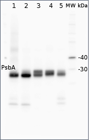
| application example 1 2 µg of total protein from (1) Arabidopsis thaliana leaf extracted with Protein ExtrationBuffer, PEB (AS08 300), (2) Hordeum vulgare leaf extracted with PEB, (3)Chlamydomonas reinhardtii total cell extracted with PEB, (4) Synechococcus sp. 7942 total cell extracted with PEB, (5) Anabaena sp. total cell extracted with PEB were separated on 4-12% NuPage (Invitrogen) LDS-PAGE and blotted 1h to PVDF. Blots were blocked immediately following transfer in 2% ECL Advance blocking reagent (GE Healthcare) in 20 mM Tris, 137 mM sodium chloride pH 7.6 with 0.1% (v/v) Tween-20 (TBS-T) for 1h at room temperature with agitation. Blots were incubated in the primary antibody at a dilution of 1: 50 000 for 1h at room temperature with agitation. The antibody solution was decanted and the blot was rinsed briefly twice, then washed once for 15 min and 3 times for 5 min in TBS-T at room temperature with agitation. Blots were incubated in secondary antibody (anti-rabbit IgG horse radish peroxidase conjugated, recommended secondary antibody AS09 602) diluted to 1:50 000 in 2% ECL Advance blocking solution for 1h at room temperature with agitation. The blots were washed as above and developed for 5 min with ECL Advance detection reagent according the manufacturers instructions. Images of the blots were obtained using a CCD imager (FluorSMax, Bio-Rad) and Quantity One software (Bio-Rad). |
 |
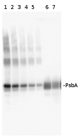
| application example 2 Varying amounts of PsbA protein standard (AS01 016S) 250 fmol (1), 125 fmol (2), 62.5 fmol (3), 31.25 fmol (4), 15.625 fmol (5) and 2 µg of total protein from Med4 (6,7)extracted with Protein Extration Buffer, PEB (AS08 300). Samples were diluted with 1X sample buffer (NuPAGE LDS sample buffer (Invitrogen) supplemented with 50 mM DTT and heat at 70°C for 5 min and keept on ice before loading. Protein samples were separated on 4-12% Bolt Plus gels, LDS-PAGE and blotted for 70 minutes to PVDF using tank transfer. Blots were blocked immediately following transfer in 2% blocking reagent (GE RPN 2125; Healthcare) or 5% non-fat milk dissolved in 20 mM Tris, 137 mM sodium chloride pH 7.6 with 0.1% (v/v) Tween-20 (TBS-T) for 1h at room temperature with agitation. Blots were incubated in the primary antibody at a dilution of 1: 10 000 (in blocking reagent) for 1h at room temperature with agitation. The antibody solution was decanted and the blot was rinsed briefly twice, and then washed 1x15 min and 3x5 min with TBS-T at room temperature with agitation. Blots were incubated in secondary antibody (goat anti-rabbit IgG horse radish peroxidase conjugated, recommended secondary antibody AS09 602, Agrisera) diluted to 1:25 000 in blocking reagent for 1h at room temperature with agitation. The blots were washed as above. The blot was developed for 5 min with TMA-6 (Lumigen) detection reagent according the manufacturers instructions. Images of the blots were obtained using a CCD imager (VersaDoc MP 4000) and Quantity One software (Bio-Rad). Exposure time was 30 seconds. |
 |
风险提示:丁香通仅作为第三方平台,为商家信息发布提供平台空间。用户咨询产品时请注意保护个人信息及财产安全,合理判断,谨慎选购商品,商家和用户对交易行为负责。对于医疗器械类产品,请先查证核实企业经营资质和医疗器械产品注册证情况。
 文献和实验
文献和实验三句话读懂一篇 CNS:喝速溶咖啡,或会缩短端粒长度;为什么女性更易患抑郁症?
启动子区的稳定而促进基因激活,而 H3K4me2/3 对转录起始没有明显的调控作用,同时 H3K4me2/3 相对于 H3K4me1 更加动态。 图 3:来源 Cell Research 4. Nature:解析首个藻胆体-光系统 II-光系统 I-捕光复合物超大复合体的结构 大多数光合蛋白复合物在细胞内的天然状态下它们之间相互作用的方式以及能量传递的途径尚不清楚。 2023 年 3 月 15 日,清华大学生命科学学院隋森芳院士等在 Nature 杂志发表研究论文 In situ
一、原理 叶绿体色素在照光时能辐射出荧光。研究叶绿体色素荧光性质,有助于了解它的分子激发态,分子之间的能量传递以及分子在活体内的排列。叶绿体光诱导荧光强度的变化(以下简称可变荧光)是由于叶绿体吸收光能后,光能在转化和电子传递过程中受阻,能量不能正常的传递下去,而以荧光的形式释放出来,使荧光的强度增加。如果这种变化是由光的影响引起的,就叫光诱导的可变荧光。当然,也可由其它条件诱导的可变荧光。在室温条件下,叶绿体在685nm处呈现一个荧光发射峰,它是由光系统II发射出来的,这部分荧光称为固定荧
10.10 Nature 子刊揭示控制自身免疫反应的分子机制
Confers Sensitivity to the MPS1 Inhibitor BOS172722 in Triple-Negative Breast Cancers ⑤ PNAS:绿藻光系统 II-捕光天线超大复合体三维结构光系统 II(PSII) 作为光合水氧化的场所,是位于光合生物类囊体膜上的一个重要蛋白质机器,对地球上生命具有重要意义。目前,在原子、分子水平上揭示光系统 II 光能捕获、传递及转化的精确机制仍存在巨大挑战。近日,中国科学院植物研究所沈建仁、匡廷云研究团队与浙江大学张兴研究团队合作
 技术资料
技术资料暂无技术资料 索取技术资料

