
大家都在搜

手机验证

询价列表

暂时没有已询价产品
![Grp78 antibody [N2C1], Internal](https://img1.dxycdn.com/2023/0711/255/2344116916935858761-14.jpg)
Grp78 antibody
[N2C1], Internal

企业认证
Recombinant protein encompassing a sequence within the center region of human Grp78. The exact sequence is proprietary.
IgG
Liquid
Store as concentrated solution. Centrifuge briefly prior to opening vial. For short-term storage (1-2 weeks), store at 4ºC. For long-term storage, aliquot and store at -20ºC or below. Avoid multiple freeze-thaw cycles.
Polyclonal
Unconjugated
Human, Mouse, Rat, Zebrafish, Mosquito
12 months from the shipping date of the product.
Human
GTX102580
Primary Antibodies
Available
GeneTex
Rabbit
WB, ICC/IF, IHC-P, IP
1.28 mg/ml (Please refer to the vial label for the specific concentration.)
Knockdown/Knockout validation was supported by customer review data.
Grp78 antibody [N2C1], Internal
Grp78 抗体 [N2C1], Internal
100 μl/25 μl
| 规格: | 100 μl | 产品价格: | ¥4000.0 |
|---|---|---|---|
| 规格: | 25 μl | 产品价格: | ¥1700.0 |

Non-transfected (–) and transfected (+) HepG2 whole cell extracts (30 μg) were separated by 7.5% SDS-PAGE, and the membrane was blotted with Grp78 antibody [N2C1], Internal (GTX102580) diluted at 1:10000. The HRP-conjugated anti-rabbit IgG antibody (GTX213110-01) was used to detect the primary antibody.

Grp78 antibody [N2C1], Internal detects Grp78 protein at cytoplasm in rat prostate by immunohistochemical analysis.
Sample: Paraffin-embedded rat prostate.
Grp78 antibody [N2C1], Internal (GTX102580) diluted at 1:500.
Antigen Retrieval: Citrate buffer, pH 6.0, 15 min

Grp78 antibody [N2C1], Internal detects Grp78 protein at cytosol on mouse intestine by immunohistochemical analysis.
Sample: Paraffin-embedded mouse intestine.
Grp78 antibody [N2C1], Internal (GTX102580) dilution: 1:500.
Antigen Retrieval: Trilogy™ (EDTA based, pH 8.0) buffer, 15min

Whole zebrafish extract (30 μg) was separated by 7.5% SDS-PAGE, and the membrane was blotted with Grp78 antibody [N2C1], Internal (GTX102580) diluted at 1:1000. The HRP-conjugated anti-rabbit IgG antibody (GTX213110-01) was used to detect the primary antibody, and the signal was developed with Trident femto Western HRP Substrate.
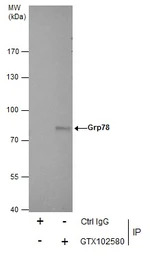
Immunoprecipitation of Grp78 protein from HepG2 whole cell extracts using 5 μg of Grp78 antibody [N2C1], Internal (GTX102580).
Western blot analysis was performed using Grp78 antibody [N2C1], Internal (GTX102580).
EasyBlot anti-Rabbit IgG (GTX221666-01) was used as a secondary reagent.
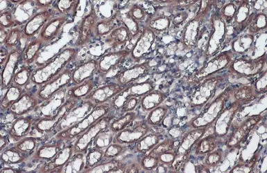
Grp78 antibody [N2C1], Internal detects Grp78 protein at cytoplasm by immunohistochemical analysis.Sample: Paraffin-embedded rat kidney.Grp78 stained by Grp78 antibody [N2C1], Internal (GTX102580) diluted at 1:1000.Antigen Retrieval: Citrate buffer, pH 6.0, 15 min
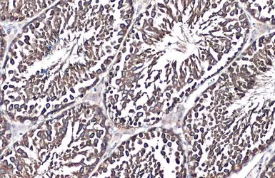
Grp78 antibody [N2C1], Internal detects Grp78 protein at cytoplasm by immunohistochemical analysis.Sample: Paraffin-embedded mouse testis.Grp78 stained by Grp78 antibody [N2C1], Internal (GTX102580) diluted at 1:500.Antigen Retrieval: Citrate buffer, pH 6.0, 15 min
Grp78 antibody [N2C1], Internal detects Grp78 protein at cytosol on rat hind brain by immunohistochemical analysis.
Sample: Paraffin-embedded rat hind brain.
Grp78 antibody [N2C1], Internal (GTX102580) dilution: 1:500.
Antigen Retrieval: Trilogy™ (EDTA based, pH 8.0) buffer, 15min
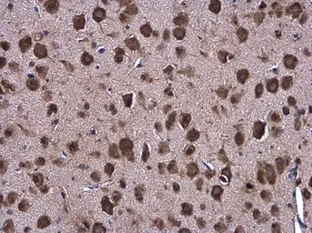
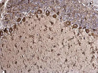
Grp78 antibody [N2C1], Internal detects Grp78 protein at cytoplasm in mouse brain by immunohistochemical analysis.
Sample: Paraffin-embedded mouse brain.
Grp78 antibody [N2C1], Internal (GTX102580) diluted at 1:500.
Antigen Retrieval: Citrate buffer, pH 6.0, 15 min
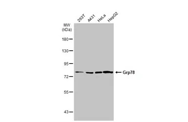
Various whole cell extracts (30 μg) were separated by 7.5% SDS-PAGE, and the membrane was blotted with Grp78 antibody [N2C1], Internal (GTX102580) diluted at 1:5000. The HRP-conjugated anti-rabbit IgG antibody (GTX213110-01) was used to detect the primary antibody.
风险提示:丁香通仅作为第三方平台,为商家信息发布提供平台空间。用户咨询产品时请注意保护个人信息及财产安全,合理判断,谨慎选购商品,商家和用户对交易行为负责。对于医疗器械类产品,请先查证核实企业经营资质和医疗器械产品注册证情况。