

- 询价
- ibidi

- 德国
- 80446
- 2026年07月10日

企业认证
相关产品推荐更多 >


万千商家帮你免费找货
0 人在求购买到急需产品
- 详细信息
- 技术资料
- 库存:
大量
- 供应商:
上海净信/拓赫机电/雷萌生物
- 现货状态:
现货
- 保修期:
1
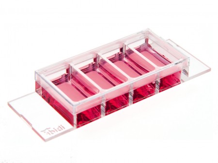
μ培养载玻片 4孔Ph+ 

一个开放的四孔培养载玻片,带有一个特殊中间板,非常适用于相差和高端的荧光显微镜
整个培养孔具有优秀的相差-没有弯月面现象
一体化小室,既可用于细胞培养,也可用于显微镜成像
使用高分辨率显微镜可以通过玻璃片底部进行样品观察
没有盖玻片,不会泄露,用于快速,简单的免疫荧光。
| 货号 | 产品名称 | 规格(个/盒) |
| 80446 | µ-Slide4孔Ph+腔室载玻片,ibiTreat底部处理 | 15 |
| 80442 | µ-Slide 4孔Ph+腔室载玻片,Collagen IV底部处理 | 15 |
| 80443 | µ-Slide 4孔Ph+腔室载玻片,Fibronectin底部处理 | 15 |
| 80444 | µ-Slide 4孔Ph+腔室载玻片,Poly-L-Lysine底部处理 | 15 |
| 80445 | µ-Slide 4孔Ph+腔室载玻片,Poly-D-Lysine底部处理 | 15 |
| 80441 | µ-Slide 4孔Ph+腔室载玻片,无包被 | 15 |
| 80447 | µ-Slide 4孔Ph+腔室载玻片,玻璃底 | 15 |
应用
细胞培养以及细胞培养物的显微镜观察。
转染分析
活细胞和固定细胞的免疫荧光显微镜观察
长时间段下活细胞成像 
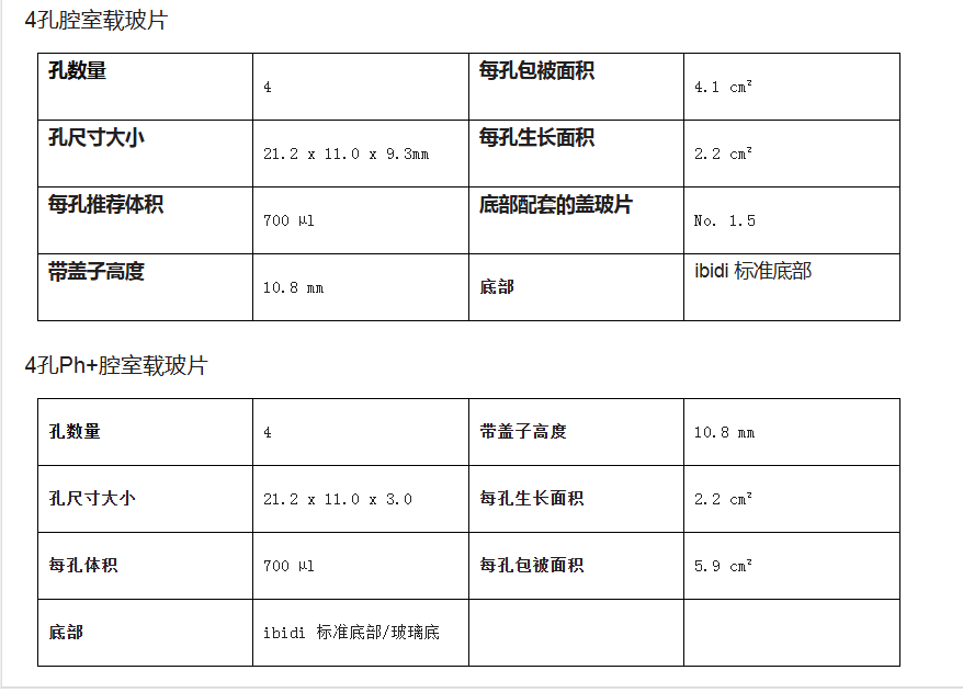
技术指标
| 孔数量 | 4 | 带盖子高度 | 10.8 mm |
| 孔尺寸大小 | 21.2 x 11.0 x 3.0 | 每孔生成面积 | 2.2 cm² |
| 每孔体积 | 700 µl | 每孔生成面积 | 5.9 cm² |
| 底部ibidi 标准底部 | |||
技术特点
开放式,两个独立的培养孔载玻片
每个培养孔上的中间板可以避免弯液面形成
从两侧缝隙很容易加液,无气泡形成,
适用于高端显微镜的优秀光学成像特质
兼容染色,固定
最佳细胞粘附的表面Ibitreat
生物兼容性塑料生产,无胶水,不泄露
Phase contrast microscopy is the most commonly used, transmitted light technique in cell culture. When working with phase contrast microscopy, it is crucial to have the two phase rings adjusted to each other. In open wells, the meniscus at the air-water-interphase works like a lens that refracts the beam path. This miscalibrates the phase rings, leading to poor contrast in the microscopic image.
U-slide 4 well U-slide 4 wellPh+ 


Working with the µ-Slide 4 well Ph+ diminishes the meniscus, so that the whole optical system is aligned, no matter which position of the well is being imaged.
Cross Section Through a Well of the µ-Slide 4 well Ph+ with a Transmitted Light Beam Path 

The illustration on the left shows the perturbing effect of a meniscus. Light is refracted on the air-water-interface, leading to poor contrast in microscopy. Only the small center part exhibits satisfying phase contrast.
Working with the µ-Slide 4 well Ph+ diminishes the meniscus and increases the area of nicely contrasted cells. This nice contrast is due to the parallel beam path that is created by the plate.
Comparison Ph+ Well versus Standard Well:
No Meniscus Effect in Ph+ Well 
µ-Slide 4 well Ph+
Excellent Phase Contrast
Excellent Fluorescence Microscopy 
tandard Well
Poor Phase Contrast
Excellent Fluorescence Microscopy
风险提示:丁香通仅作为第三方平台,为商家信息发布提供平台空间。用户咨询产品时请注意保护个人信息及财产安全,合理判断,谨慎选购商品,商家和用户对交易行为负责。对于医疗器械类产品,请先查证核实企业经营资质和医疗器械产品注册证情况。
 技术资料
技术资料需要更多技术资料 索取更多技术资料
资料下载:
80446说明.pdf 附 (下载 0 次)
80446实验 (1).pdf 附 (下载 0 次)
80446说明2.pdf 附 (下载 0 次)
80446说明4.pdf 附 (下载 0 次)
80446说明3.pdf 附 (下载 0 次)
80446实验.pdf 附 (下载 0 次)
请 [登录] 后再下载!

