产品描述:H2AX Rabbit Polyclonal Antibody
别名:H2AX_HUMAN; 2A.X variant histone; H2A.X; H2A/X; H2AFX; Histone H2AX;
免疫原:KLH conjugated synthetic peptide derived from human H2AX (30-143/143aa)
预测反应性:Bovine, Canine, Equine, Porcine, Rabbit
分子量:16 kDa
应用稀释比例:WB=1:500-2000, IHC-P=1:100-500, IHC-F=1:400-800, IF=1:100-500
防腐剂:0.01M TBS (pH7.4) with 1% rAlbumin, 0.02% Proclin300 and 50% Glycerol.
纯化:Affinity purified by Protein A
保存说明:Maintain refrigerated at 2-8°C for up to 2 weeks. For long term storage store at -20°C in small aliquots to prevent freeze-thaw cycles.
UniProt ID:P16104
Note:For research use only.
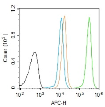
Blank control (Black line): Molt4 (Black). Primary Antibody (green line): Rabbit Anti-Histone H2A.x antibody (orb13454), Dilution: 1 µg/10^6 cells, Isotype Control Antibody (orange line): Rabbit IgG. Secondary Antibody (white blue line): Goat anti-rabbit IgG-AF647, Dilution: 1 µg/Test. Protocol, The cells were fixed with 4% PFA (10 min at room temperature) and then permeabilized with 90% ice-cold methanol for 20 min at room temperature. The cells were then incubated in 5% BSA to block non-specific protein-protein interactions for 30 min at room temperature. Cells stained with Primary Antibody for 30 min at room temperature. The secondary antibody used for 40 min at room temperature. Acquisition of 20000 events was performed.
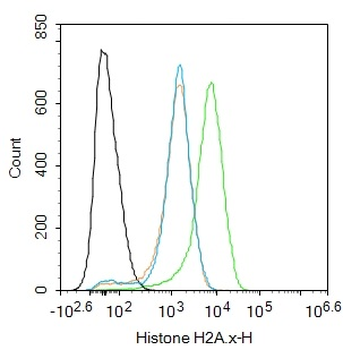
Blank control: Jurkat. Primary Antibody (green line): Rabbit Anti-Histone H2A.x antibody (orb13454), Dilution: 1 ug/Test, Secondary Antibody: Goat anti-rabbit IgG-FITC, Dilution: 0.5 ug/Test. Protocol, The cells were fixed with 4% PFA (10 min at room temperature) and then permeabilized with 90% ice-cold methanol for 20 min at -20°C. The cells were then incubated in 5% BSA to block non-specific protein-protein interactions for 30 min at room temperature. Cells stained with Primary Antibody for 30 min at room temperature. The secondary antibody used for 40 min at room temperature. Acquisition of 20000 events was performed.
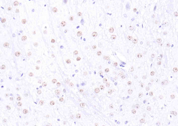
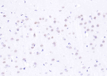
Fixative-fixed, paraffin embedded (mouse brain), Antigen retrieval by boiling in sodium citrate buffer (pH6.0) for 15 min, Block endogenous peroxidase by 3% hydrogen peroxide for 20 minutes, Blocking buffer (normal goat serum) at 37°C for 30 min, Antibody incubation with (H2AX) Polyclonal Antibody, Unconjugated at 1:200 overnight at 4°C, followed by operating according to SP Kit (Rabbit) instructionsand DAB staining.
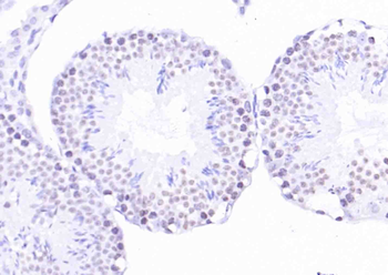
Fixative-fixed, paraffin embedded (mouse testis), Antigen retrieval by boiling in sodium citrate buffer (pH6.0) for 15 min, Block endogenous peroxidase by 3% hydrogen peroxide for 20 minutes, Blocking buffer (normal goat serum) at 37°C for 30 min, Antibody incubation with (H2AX) Polyclonal Antibody, Unconjugated (orb13454) at 1:200 overnight at 4°C, followed by operating according to SP Kit (Rabbit) instructionsand DAB staining.
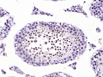
Fixative-fixed, paraffin embedded (mouse testis), Antigen retrieval by boiling in sodium citrate buffer (pH6.0) for 15 min, Block endogenous peroxidase by 3% hydrogen peroxide for 20 minutes, Blocking buffer (normal goat serum) at 37°C for 30 min, Antibody incubation with (Histone H2A.x) Polyclonal Antibody, Unconjugated (orb13454) at 1:200 overnight at 4°C, followed by operating according to SP Kit (Rabbit) instructionsand DAB staining.
Fixative-fixed, paraffin embedded (rat brain), Antigen retrieval by boiling in sodium citrate buffer (pH6.0) for 15 min, Block endogenous peroxidase by 3% hydrogen peroxide for 20 minutes, Blocking buffer (normal goat serum) at 37°C for 30 min, Antibody incubation with (H2AX) Polyclonal Antibody, Unconjugated (orb13454) at 1:200 overnight at 4°C, followed by operating according to SP Kit (Rabbit) instructionsand DAB staining.
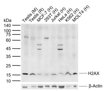
Sample: Lane 1: Mouse Testis tissue lysates, Lane 2: Rat Testis tissue lysates, Lane 3: Human MCF-7 cell lysates, Lane 4: Human 293T cell lysates, Lane 5: Human Jurkat cell lysates, Lane 6: Human HeLa cell lysates, Lane 7: Human K562 cell lysates, Lane 8: Human MOLT4 cell lysates, Primary: Anti-H2AX (orb13454) at 1/1000 dilution, Secondary: IRDye800CW Goat Anti-Rabbit IgG at 1/20000 dilution, Predicted band size: 16 kDa, Observed band size: 16 kDa.
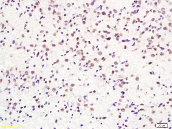
Tissue/Cell: human glioma tissue, 4% Fixative-fixed and paraffin-embedded, Antigen retrieval: citrate buffer (0.01M, pH 6.0), Boiling bathing for 15 min, Block endogenous peroxidase by 3% Hydrogen peroxide for 30 min, Blocking buffer (normal goat serum) at 37°C for 20 min, Incubation: Anti-H2AX/Histone H2A.x Polyclonal Antibody, Unconjugated (orb13454) 1:200, overnight at 4°C, followed by conjugation to the secondary antibody and DAB staining.


 文献和实验
文献和实验 技术资料
技术资料
