

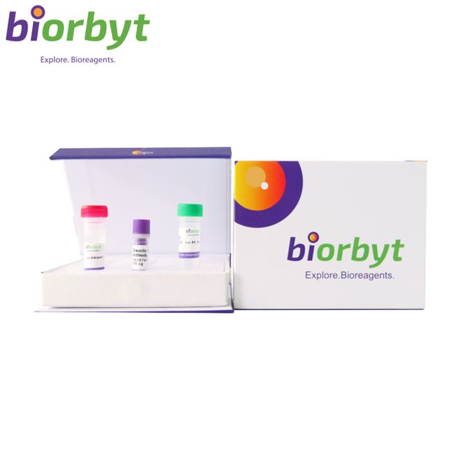
- ¥7488
- Biorbyt

- orb151518
- 英国
- 2026年07月29日
- FC, ICC, IF, IHC, IP, WB
- Rabbit
- Bovine, Canine, Gallus, Guinea pig, Hamster, Human, Monkey, Mouse, Porcine, Rabbit, Rat, Sheep

企业认证
相关产品推荐更多 >


万千商家帮你免费找货
0 人在求购买到急需产品
相关阅读
源叶生物 十万种现货试剂速发,品质稳、品类全,实验不再等!Takara 秋促来袭,提取+反转录+定量套装仅 1200 元abinScience 体内级抗体 全年锁价 低至 ¥1200阿拉丁 22万+ 科研级产品现货速发,品质严控、科研无忧- 详细信息
- 文献和实验
- 技术资料
- 抗体名:
Calreticulin Antibody (RPE) 抗体
- 抗体英文名:
Calreticulin Antibody (RPE)
- 靶点:
Calreticulin
- 浓度:
1 mg/ml
- 应用范围:
FC, ICC, IF, IHC, IP, WB
- 宿主:
Rabbit
- 适应物种:
Bovine, Canine, Gallus, Guinea pig, Hamster, Human, Monkey, Mouse, Porcine, Rabbit, Rat, Sheep
- 保质期:
12 months from date of receipt.
- 抗原来源:
详询
- 目录编号:
orb151518
- 级别:
科研
- 库存:
99
- 供应商:
Biorbyt
- 标记物:
RPE
- 克隆性:
Polyclonal
- 保存条件:
详询
- 免疫原:
详询
- 规格:
200 ul
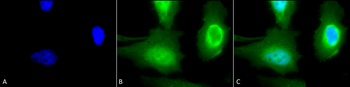
描述: Rabbit polyclonal to Calreticulin (RPE). Calreticulin is a multifunctional, highly conserved Ca2+ -binding protein that is localized to the endoplasmic reticulum (ER), but has also been detected in the nucleus and nuclear envelop. Like many other ER proteins, it has the conserved ER retention KDEL (Lys-Asp-Glu-Leu) sequence at its C-terminus (1-3). CRTs three domains include a 180 residue N-terminal domain, a proline-rich P-domain (residues 189-288) that binds Ca2+ with high affinity and shares homology with calnexin (CNX) and calmegin, and a 110 residue C-terminal domain that binds Ca2+ with low affinity but high capacity (1,3). Recent studies suggest that this soluble ER protein has a multifunctional role. It appears to be involved in calcium storage and regulation as well as having a molecular chaperone activity. It has been shown to interact with the cytoskeleton and to be involved in the regulation of gene expression. Calreticulin may also play a role in cellular proliferation including its apparent activity in the proliferation of certain viruses within mammalian host cells , and it has also been shown to be induced in response to various types of cell stress including amino acid deprivation. Close interconnections among protein synthesis, gene expression and calcium signaling have been observed by many researchers in recent years. Calreticulin might be centrally located and therefore it crucially participates in the coordination of many functions by the cell. Studies also suggest its involvement in a few diseases such as systemic lupus erythematosus, rheumatoid arthritis, celiac disease, complete congenital heart block, and halothane hepatitis..
别名: Calreticulin, CALR, CALR_HUMAN, Calregulin, cC1qR, CRP55, ERp60, HSCBP, RO, SSA, grp60
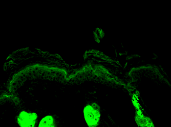
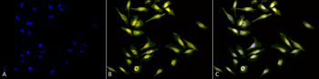
应用稀释比例: WB (1:1000), IHC (1:100), ICC/IF (1:100)
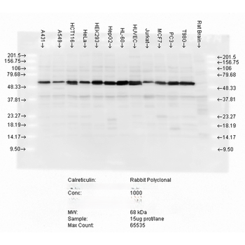
应用说明: A 1:1000 dilution was sufficient for detection of Calreticulin in 20 µg of HeLa cell lysate by ECL immunoblot analysis.
免疫原: Human calreticulin synthetic peptide with a cysteine residue added and the peptide conjugated to KLH
分子量: 63kDa
防腐剂: 95.46mM Phosphate, 2.48mM MES and 2mM EDTA
纯化方法: Peptide Affinity Purified
特异性: Detects ~63kDa.
研究领域: Metabolism Research
UniProt ID: P27797
NCBI 参考序列: NP_004334.1
Entrez 基因 ID: 811
保存条件: Conjugated antibodies should be stored according to the product label
备注: For research use only.
风险提示:丁香通仅作为第三方平台,为商家信息发布提供平台空间。用户咨询产品时请注意保护个人信息及财产安全,合理判断,谨慎选购商品,商家和用户对交易行为负责。对于医疗器械类产品,请先查证核实企业经营资质和医疗器械产品注册证情况。
 文献和实验
different tissues of the eye, i.e., cornea, lens, iris, retina, optic nerve, retinal pigment epithelium (RPE), and sclera. We further describe in detail how to process these eye samples in order to obtain high?quality RNA for RNA expression profiling studies
基本没有RPE原有性质特征,只具有成纤维细胞的特征。 【qkk1229】同意N代后就是成纤维细胞的模样,很多细胞体外培养到后来,都长的象成纤维细胞,不知道为什么,有人知道吗? 【jamesxia】去除成纤维细胞 1、反复贴壁法(差速贴壁法):倒去旧培养液,用Hank′s液洗3次,然后加入消化液(0.2%EDTA+0.25%胰蛋白酶),37℃10min,镜下可见成纤维细胞成圆形,癌细胞维持原状。吸出消化液,加Hank′s液吹打细胞使成纤维细胞脱落,吸出旧液,组织块和癌细胞团用Hank′s液轻轻
 技术资料
暂无技术资料 索取技术资料