

- 询价
- Agrisera
- AS10 710
- 瑞典
- 2026年02月06日
- ChIp-qPCR (ChIp-qPCR), Immunocytochemistry (ICC), Immunofluorescence (IF), Western blot (WB)
- Rabbit
- Arabidopsis thaliana, Brassica oleracea, Capsicum annuum, Cicer arietinum L., Chlamydomonas acidophila, Chlamydomonas reinhardtii, Cucumis sativus L cv Suyo, human, Nicotiana benthamiana, Physcomitrella patens, Salicornia europaea, Solanum lycopersicum, S

企业认证
相关产品推荐更多 >

万千商家帮你免费找货
0 人在求购买到急需产品
- 详细信息
- 文献和实验
- 技术资料
- 抗体名:
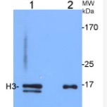
H3 | Histone H3 (rabbit antibody) (nuclear marker)
- 抗体英文名:
H3 | Histone H3 (rabbit antibody) (nuclear marker)
- 靶点:
Histone 3 (H3) located in nuclei, incorporated into chromatin. Present in nucleosome together with H2A, H2B and H4.
- 应用范围:
ChIp-qPCR (ChIp-qPCR), Immunocytochemistry (ICC), Immunofluorescence (IF), Western blot (WB)
- 宿主:
Rabbit
- 适应物种:
Arabidopsis thaliana, Brassica oleracea, Capsicum annuum, Cicer arietinum L., Chlamydomonas acidophila, Chlamydomonas reinhardtii, Cucumis sativus L cv Suyo, human, Nicotiana benthamiana, Physcomitrella patens, Salicornia europaea, Solanum lycopersicum, S
- 抗原来源:
P59169 , P59226 , Q9FXI7
- 级别:
Serum
- 供应商:
Agrisera AB
- 克隆性:
单克隆
- 保存条件:
Store lyophilized/reconstituted at -20°C; once reconstituted make aliquots to avoid repeated freeze-thaw cycles. Please, remember to spin tubes briefly prior to opening them to avoid any losses that might occur from lyophilized material adhering to the ca
- 形态:
Lyophilized
- 免疫原:
KLH-conjugated synthetic peptide derived from known H3 sequences, inluding Arabidopsis thaliana H3.3 P59169 (At4g40030, At4g40040, At5g10980), H3.2 P59226 (At1g09200, At3g27360, At5g10390, At5g10400, At5g65360), H3-like 2 Q9FXI7 (At1g19890)
- 规格:
50 µl
风险提示:丁香通仅作为第三方平台,为商家信息发布提供平台空间。用户咨询产品时请注意保护个人信息及财产安全,合理判断,谨慎选购商品,商家和用户对交易行为负责。对于医疗器械类产品,请先查证核实企业经营资质和医疗器械产品注册证情况。
 文献和实验
文献和实验Detection of Histone H3 Phosphorylation in Cultured Cells and Tissue Sections by Immunostaining
. The protocol described here allows the detection of phosphorylated histones in tissue-cultured cells and tissue sections by fluorescent or bright-field immunostaining analysis. Here we used a serine 10 specific P-histone H3 antibody to determine
FACS-Based Detection of Phosphorylated Histone H3 for the Quantitation of Mitotic Cells
scanner (FACS) is described, based on the presence of an intranuclear antigen present only in mitotic cells, detected using a specific, commercially available antibody. Cell staining and FACS analysis can be done in a single day, making this a rapid
Genome-Wide Measurement of Histone H3 Replacement Dynamics in Yeast
chromatin. Understanding the dynamic behavior of chromatin is of great interest for fields ranging from transcriptional regulation through meiosis and gametogenesis. Here, we describe a protocol for measuring histone replacement rates genome wide


