

- ¥3770
- biorbyt

- orb1239340
- 英国
- 2025年12月08日
- ELISA, ICC, IF, IHC-P, WB
- Human, Mouse, Rat

企业认证
相关产品推荐更多 >


万千商家帮你免费找货
0 人在求购买到急需产品
- 详细信息
- 技术资料
- 抗体名:
DR4 Antibody抗体
- 抗体英文名:
DR4 Antibody
- 靶点:
TNFRSF10A
- 浓度:
1 mg/mL
- 应用范围:
ELISA, ICC, IF, IHC-P, WB
- 适应物种:
Human, Mouse, Rat
- 保质期:
6-12个月
- 抗原来源:
详询
- 目录编号:
orb1239340
- 级别:
科研级
- 库存:
88
- 供应商:
biorbyt
- 标记物:
Unconjugated
- 克隆性:
Polyclonal
- 形态:
Liquid
- 亚型:
IgG
- 免疫原:
Anti-DR4 antibody (orb1239340) was raised against a peptide corresponding to 19 amino acids near the carboxy terminus of human DR4. The immunogen is located within the last 50 amino acids of DR4.
- 规格:
0.02 mg
产品描述:DR4 Antibody
别名:DR4 Antibody: DR4, APO2, CD261, TRAILR1, TRAILR-1, DR4, Tumor necrosis factor receptor superfamily member 10A, Death receptor 4, TRAIL receptor 1
免疫原:Anti-DR4 antibody (orb1239340) was raised against a peptide corresponding to 19 amino acids near the carboxy terminus of human DR4. The 免疫原 is located within the last 50 amino acids of DR4.
分子量:Predicted: 50kDObserved: 55kD (Post-modification: 1 N-linked glycosylation)
防腐剂:DR4 Antibody is supplied in PBS containing 0.02% sodium azide.
纯化:DR4 Antibody is Antibody is affinity chromatography purified via peptide column.
保存说明:Maintain refrigerated at 2-8°C for up to 2 weeks. For long term storage store at -20°C in small aliquots to prevent freeze-thaw cycles.
NCBI:AAC51226
UniProt ID:O00220
Note:For research use only.
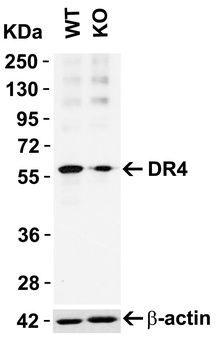
KO Validation in HeLa Cells. Loading: 10 µg of HeLa WT cell lysates or DR4 KO cell lysates. Antibodies: DR4 orb1239340 (1 µg/mL) and beta-actin orb1240312 (1 µg/mL), 1 h incubation at RT in 5% NFDM/TBST. Secondary: Goat Anti-Rabbit IgG HRP conjugate at 1:10000 dilution.
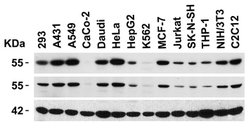
Independent Antibody Validation (IAV) via Protein Expression Profile in Cell Lines. Loading: 15 µg of lysates per lane. Antibodies: DR4 orb1239340 (1 µg/mL), DR4 orb1239343 (4 µg/mL), and beta-actin (1 µg/mL), 1h incubation at RT in 5% NFDM/TBST. Secondary: Goat anti-rabbit IgG HRP conjugate at 1:10000 dilution.
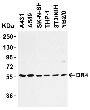
Western Blot Validation in Cell Lines. Loading: 15 µg of cell lysates per lane. Antibodies: DR4 orb1239340 (1 µg/mL), 1h incubation at RT in 5% NFDM/TBST. Secondary: Goat anti-rabbit IgG HRP conjugate at 1:10000 dilution.
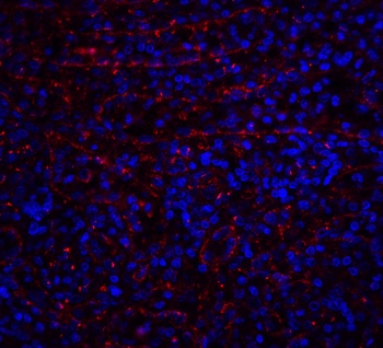
Immunofluorescence Validation of DR4. Immunofluorescent analysis of 4% Fixative-fixed human spleen tissue labeling DR4 with orb1239340 at 20 µg/mL, followed by goat anti-rabbit IgG secondary antibody at 1/500 dilution (red) and DAPI staining (blue). Image showing membrane staining on human spleen cells.
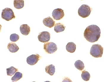
Immunocytochemistry Validation of DR4 in HeLa Cells. Immunocytochemical analysis of HeLa cells using anti-DR4 antibody (orb1239340) at 10 µg/ml. Cells was fixed with formaldehyde and blocked with 10% serum for 1 h at RT; antigen retrieval was by heat mediation with a citrate buffer (pH6). Samples were incubated with primary antibody overnight at 4°C. A goat anti-rabbit IgG H&L (HRP) at 1/250 was used as secondary. Counter stained with Hematoxylin.
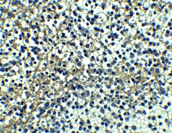
Immunohistochemistry Validation of DR4. Immunohistochemical analysis of paraffin-embedded human spleen tissue using anti-DR4 antibody (orb1239340) at 10 µg/ml. Tissue was fixed with formaldehyde and blocked with 10% serum for 1 h at RT; antigen retrieval was by heat mediation with a citrate buffer (pH6). Samples were incubated with primary antibody overnight at 4°C. A goat anti-rabbit IgG H&L (HRP) at 1/250 was used as secondary. Counter stained with Hematoxylin.
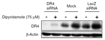
KD Validation in SW480 cells (Goda et al., 2008). The expression of DR4 was knocked down via DR4 siRNA, 24 h latercells were treated with dipyridamole for 24 h. DR4 protein expression detected by anti-DR4 antibodies (orb1239340) was disrupted. Dipyridamole up-regulated the expression of DR4.
Immunofluorescence Validation of DR4 in rat brain (Cantarella et al., 2014). DR4 protein expression detected by anti-DR4 antibodies (orb1239340) was increased after transient brain ischemia (tMCAO) and decreased after pre-conditioning stimulus. Confocal microscopic images displaying NeuN (a, d, g) (green), DR4 (b, e, h) (red), and Merge (c, f, i) (yellow) in the brain peri-ischemic region of rats after 5 h.
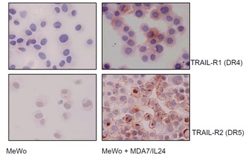
Immunocytochemistry Validation of DR4 in human melanoma cells (Ekmekcioglu et al., 2008). MeWo melanoma cells were exposed to affinity-purified MDA7/IL-24. After 48 h of treatment, cells were collected and cytospins prepared for cytochemical assessment of their TRAIL receptor (R1 and R2) expression (anti-DR4 (orb1239340) or anti-DR5, AEC, hematoxylin). Both DR4 and DR5 expression were upregulated in MeWo cells after treatment.
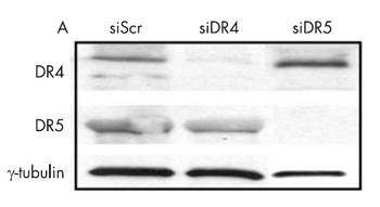
KD Validation in Huh7 cells (Malhi et al., 2007). Western blot analysis with anti-DR4 antibodies (orb1239340) was performed for DR5 and DR4 expression using whole cell lysates from Huh 7 cells transfected with respective siRNAs. In cells treated with siDR4, a decrease in DR4 level was observed, DR5 levels were unchanged. Scrambled siRNA was used as control.
风险提示:丁香通仅作为第三方平台,为商家信息发布提供平台空间。用户咨询产品时请注意保护个人信息及财产安全,合理判断,谨慎选购商品,商家和用户对交易行为负责。对于医疗器械类产品,请先查证核实企业经营资质和医疗器械产品注册证情况。
 技术资料
技术资料暂无技术资料 索取技术资料
DR4 Antibody抗体,orb1239340,biorbyt
¥3770
