

- ¥8320
- biorbyt

- orb151949
- 英国
- 2025年12月04日
- ELISA, ICC, IF, WB
- Gallus
- Human, Mouse, Rat

企业认证
相关产品推荐更多 >
Cyclin D2 (phospho Thr280) rabbit pAb抗体,orb770787,biorbyt
¥3250
Glutamine Synthetase antibody 抗体,orb2276275,Biorbyt
¥9464
Phospho-Glucocorticoid Receptor (Ser211) Antibody (Cy5.5) 抗体,orb1581701,Biorbyt
¥4796
G3BP2 Antibody - middle region : HRP 抗体,orb2083658,Biorbyt
¥8216
Anti-CES1P1 Antibody 抗体,orb1880929,Biorbyt
¥2299
万千商家帮你免费找货
0 人在求购买到急需产品
- 详细信息
- 文献和实验
- 技术资料
- 抗体名:
HSP70 Antibody (HRP)抗体
- 抗体英文名:
HSP70 Antibody (HRP)
- 靶点:
HSP70
- 浓度:
1 mg/ml
- 应用范围:
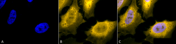
ELISA, ICC, IF, WB
- 宿主:
Gallus
- 适应物种:
Human, Mouse, Rat
- 保质期:
6-12个月
- 抗原来源:
详询
- 目录编号:
orb151949
- 级别:
科研级
- 库存:
88
- 供应商:
biorbyt
- 标记物:
HRP
- 克隆性:
Polyclonal
- 亚型:
IgY
- 免疫原:
Full length Human HSP70
- 规格:
100 ug
别名:HSPA1A, HSPA1B, HSPA1, HSP70, HSP70-1, HSP70.1, HSP70-2, HSP72, HSP73, HSX70, Heat shock 70 kDa protein 1A, Heat shock 70 kDa protein 1B
免疫原:Full length Human HSP70
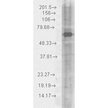
分子量:70kDa
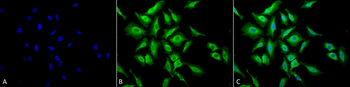
应用稀释比例:WB (1:1000), ICC/IF (1:120)
应用注释:1 µg/ml of SMC-178 was sufficient for detection of HSP70 in 20 µg of heat shocked HeLa cell lysate by colorimetric immunoblot analysis using Goat anti-chicken IgY:HRP as the secondary antibody.
防腐剂:73.64mM Carbonate, 54.55mM Ethanolamine, 45.45mM Cyanoborohydride, 18.18mM Sodium Hydroxide, 0.23mM Citrate
纯化:PEG Purified
保存说明:Conjugated antibodies should be stored according to the product label
NCBI:NP_005336.3
Entrez:3303
UniProt ID:P0DMV8, P0DMV9
Note:For research use only.
风险提示:丁香通仅作为第三方平台,为商家信息发布提供平台空间。用户咨询产品时请注意保护个人信息及财产安全,合理判断,谨慎选购商品,商家和用户对交易行为负责。对于医疗器械类产品,请先查证核实企业经营资质和医疗器械产品注册证情况。
 文献和实验
文献和实验Conjugation of Antibody to HRP
of Maleimide:HRP Weigh out 1.067 mg HRP for each mg of thiolated antibody. Add 1.1 mg Sulfo-SMCC per mg of HRP Add PBS to a final concentration of 8 mg/ml HRP Mix for 2 hours on a rotator. Desalt the HRP on PBS equilibrated column. Collect
Conjugation of Antibody to HRP
Conjugation of Antibody to HRP Author: Nanci Donacki Source: Contributed by Nanci Donacki Reagents
Monoclonal Antibody Production Protocol
- PBS + 0.1% Tween 20 or TBS + 0.1% Tween 20. - Blocking solution = 2% BSA (type V ) in PBS. (Add 0.02% azide for longer storage.) - Elisa buffer = 2% BSA + 0.1% Tween 20 in PBS ( azide optional ). - Enzyme linked antibody = Horseradish peroxidase
 技术资料
技术资料暂无技术资料 索取技术资料

