产品描述:Mouse monoclonal to Nav1 (FITC). 7. Ion channels are integral membrane proteins that help establish and control the small voltage gradient across the plasma membrane of living cells by allowing the flow of ions down their electrochemical gradient. They are present in the membranes that surround all biological cells because their main function is to regulate the flow of ions across this membrane. Whereas some ion channels permit the passage of ions based on charge, others conduct based on a ionic species, such as sodium or potassium. Furthermore, in some ion channels, the passage is governed by a gate which is controlled by chemical or electrical signals, temperature, or mechanical forces. There are a few main classifications of gated ion channels. There are voltage- gated ion channels, ligand- gated, other gating systems and finally those that are classified differently, having more exotic characteristics. The first are voltage- gated ion channels which open and close in response to membrane potential. These are then separated into sodium, calcium, potassium, proton, transient receptor, and cyclic nucleotide-gated channels; each of which is responsible for a unique role. Ligand-gated ion channels are also known as ionotropic receptors, and they open in response to specific ligand molecules binding to the extracellular domain of the receptor protein. The other gated classifications include activation and inactivation by second messengers, inward-rectifier potassium channels, calcium-activated potassium channels, two-pore-domain potassium channels, light-gated channels, mechano-sensitive ion channels and cyclic nucleotide-gated channels. Finally, the other classifications are based on less normal characteristics such as two-pore channels, and transient receptor potential channels. Nav1. 7 is a voltage-gated sodium channel and plays a critical role in the generation and conduction of action potentials and is thus important for electrical signaling by most excitable cells. Therapeutically, the association of pain insensitivity with the loss of function of a certain sodium channel may have implications. Since Nav1. 7 is not present in cardiac muscle or neurons in the central nervous system, blockers of Nav1. 7 will not have direct action on these cells and thus can have less side effects than current pain medications. By performing more studies, there is a possibility to develop a new generation of drugs that can reduce the pain intensity in animals (3-5)..
别名:Nav1.7, SCN9A, ETHA, hNE Na, NE NA, PN1, Voltage gated sodium channel subunit alpha Nav1, Peripheral sodium channel 1, Neuroendocrine sodium channel
免疫原:Fusion protein amino acids 1751-1946 (C-terminus) of human Nav1.7
克隆性:N68/6 (Formerly sold as S68-6)
分子量:230kDa
应用稀释比例:WB (1:1000), IHC (1:1000), ICC/IF (1:100)
应用注释:1 µg/ml of SMC-314 was sufficient for detection of Nav1.7 in 10 µg of HEK-293 cell lysate transiently expressing Nav1.7 by colorimetric immunoblot analysis using Goat anti-mouse IgG:HRP as the secondary antibody.
防腐剂:640.91mM DMSO, 136.36 mM Ethanolamine, 126.89 mM chlorides, 9.09mM phosphates, 9.09mM NaHCO3
纯化:Protein G Purified
保存说明:Conjugated antibodies should be stored according to the product label
NCBI:NP_002968.1
Entrez:6335
UniProt ID:Q15858
Note:For research use only.
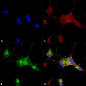
Immunocytochemistry/Immunofluorescence analysis using Mouse Anti-Nav1.7 Monoclonal Antibody, Clone N68/6. Tissue: Neuroblastoma cells (SH-SY5Y). Species: Human. Fixation: 4% PFA for 15 min. Primary Antibody: Mouse Anti-Nav1.7 Monoclonal Antibody at 1:100 for overnight at 4°C with slow rocking. Secondary Antibody: AlexaFluor 488 at 1:1000 for 1 hour at RT. Counterstain: Phalloidin-iFluor 647 (red) F-Actin stain; Hoechst (blue) nuclear stain at 1:800, 1.6mM for 20 min at RT. (A) Hoechst (blue) nuclear stain. (B) Phalloidin-iFluor 647 (red) F-Actin stain. (C) Nav1.7 Antibody (D) Composite.
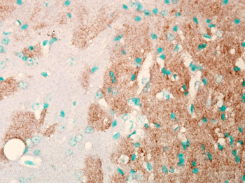
Immunohistochemistry analysis using Mouse Anti-Nav1.7 Sodium Channel Monoclonal Antibody, Clone N68/6. Tissue: Brain Slice. Species: Mouse. Fixation: Frozen sections. Primary Antibody: Mouse Anti-Nav1.7 Sodium Channel Monoclonal Antibody at 1:1000. Secondary Antibody: HRP/DAB Detection System: Biotinylated Goat Anti-Mouse, Streptavidin Peroxidase, DAB Chromogen (brown). Counterstain: Mayer Hematoxylin (purple/blue) nuclear stain.
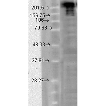
Western Blot analysis of Hamster CHO cells showing detection of Nav1.7 Sodium Channel protein using Mouse Anti-Nav1.7 Sodium Channel Monoclonal Antibody, Clone N68/6. Load: 15 μg. Block: 1.5% BSA for 30 minutes at RT. Primary Antibody: Mouse Anti-Nav1.7 Sodium Channel Monoclonal Antibody at 1:1000 for 2 hours at RT. Secondary Antibody: Sheep Anti-Mouse IgG: HRP for 1 hour at RT.
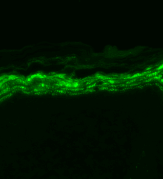
Immunohistochemistry analysis using Mouse Anti-Nav1.7 Sodium Channel Monoclonal Antibody, Clone N68/6. Tissue: backskin. Species: Mouse. Fixation: Bouin's Fixative and paraffin-embedded. Primary Antibody: Mouse Anti-Nav1.7 Sodium Channel Monoclonal Antibody at 1:100 for 1 hour at RT. Secondary Antibody: FITC Goat Anti-Mouse (green) at 1:50 for 1 hour at RT.
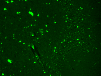
Immunohistochemistry analysis using Mouse Anti-Nav1.7 Sodium Channel Monoclonal Antibody, Clone N68/6. Tissue: hippocampus. Species: Human. Fixation: Bouin's Fixative and paraffin-embedded. Primary Antibody: Mouse Anti-Nav1.7 Sodium Channel Monoclonal Antibody at 1:1000 for 1 hour at RT. Secondary Antibody: FITC Goat Anti-Mouse (green) at 1:50 for 1 hour at RT.


 文献和实验
文献和实验 技术资料
技术资料
