


- ¥6000
- 苏州艾洛蒙
- ARD1103
- 苏州
- 2026年05月29日
- ELISA, FACS, Functional assay

企业认证
相关产品推荐更多 >


万千商家帮你免费找货
0 人在求购买到急需产品
- 详细信息
- 文献和实验
- 技术资料
- 抗体名:
抗人 CDH3 抗体 FF-21101
- 抗体英文名:
Anti-CDH3/P-cadherin Antibody(FF-21101)
- 靶点:
human CDH3
- 应用范围:
ELISA, FACS, Functional assay
- 供应商:
苏州艾洛蒙
- 库存:
100
- 级别:
研究级
- 克隆性:
单克隆
- 保存条件:
store at -20°C
- 形态:
液体
- 亚型:
Human IgG1, κ
- 规格:
1mg
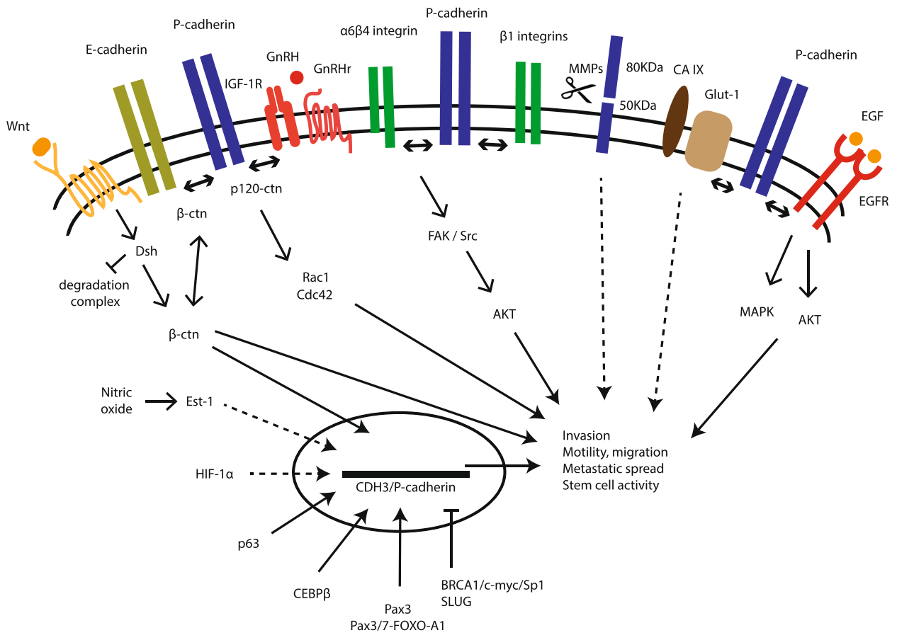
FF-21101 is a human IgG1 monoclonal antibody (mAb) targeting CDH3/P-cadherin。A radioimmunoconjugate consisting of a chimeric monoclonal antibody targeting human cadherin-3 (CDH3) and labeled, via the macrocyclic chelator 1,4,7,10-tetraazacyclododecane-1,4,7,10-tetraacetic acid (DOTA), with the beta-emitting radioisotope yttrium Y 90, with potential antineoplastic activities. Upon administration, the antibody moiety of yttrium Y 90 anti-CDH3 monoclonal antibody FF-21101 binds to CDH3 expressed on tumor cells, thereby specifically delivering cytotoxic beta radiation to CDH3-expressing tumor cells. CDH3, also known as P-cadherin, is a tumor-associated antigen (TAA) and member of the cadherin family; it is overexpressed in a variety of tumors and plays a role in cell adhesion, motility, invasion, and proliferation.
P-cadherin is overexpressed in various cancers and can be a target for radioimmunotherapy. We investigated the preclinical pharmacokinetics and pharmacology of FF-21101, an indium-111 (111In)- or yttrium-90 (90Y)-conjugated monoclonal antibody against P-cadherin, to evaluate its clinical applications. Methods: The radiochemical purity, binding affinity, and in vitro serum stability of 111In/90Y-labeled FF-21101 were evaluated. The pharmacokinetics of 111In/90Y-FF-21101 were compared in normal mice. Tumor accumulation after 111In-FF-21101 administration was investigated in mice bearing subcutaneous tumors with high (NCI-H1373), moderate (EBC-1), and negative (A549) P-cadherin expression. The tumor suppression effect following a single intravenous injection of 90Y-FF-21101 was assessed in NCI-H1373 and EBC-1 mouse xenograft models. The relationship between antibody dose and tumor accumulation was investigated in the NCI-H1373 mouse xenograft model. The radiation-absorbed dose in humans after injection of 90Y-FF-21101 was estimated using gamma camera images of cynomolgus monkeys.
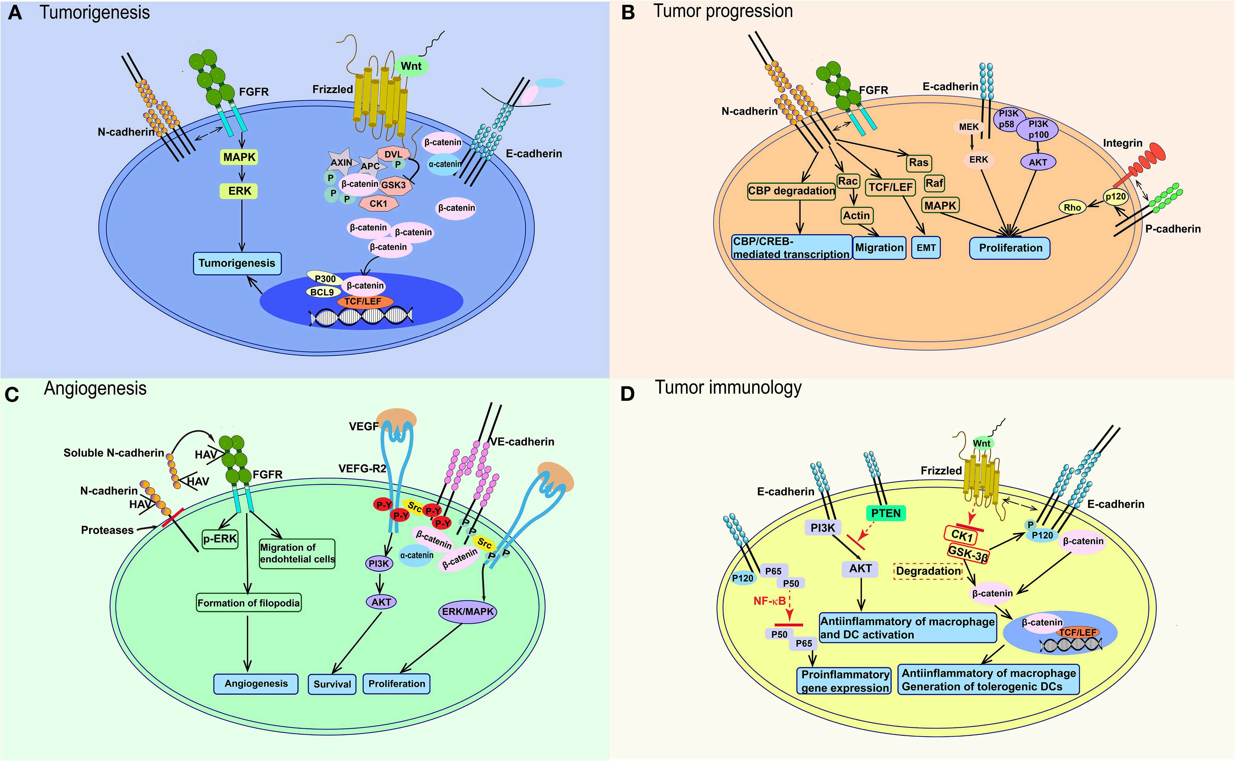
Signaling pathways participating in the function of cadherins in cancer. (A) E-cadherin loss lead to the upregulation of β-catenin in cytoplasm. After Wnt binds to Frizzled, it blocks the effects of CK1α and GSK3 on β-catenin and cause the accumulation of β-catenin in cytoplasm and nucleus, which activates TCF/LEF and co-activators. N-cadherin could affect tumorigenesis through interacting with FGFR and stimulating MAPK/ERK pathway. (B) Wnt-β-catenin pathway also works in tumor progression like in (A). E-cadherin could also activate PI3K-AKT and MEK-ERK pathways. N-cadherin activates Ras-MAPK pathway, TCF/LEF transcription factor, etc. P-cadherin could increase tumor migration through interacting with integrin. (C) In endothelial cells, VEGF-VEGFR2 activates VE-cadherin and Src, then leads to VE-cadherin phosphorylation on tyrosines, which would promote endothelial cell proliferation through PI3K/AKT signaling. ERK/MAPK signaling pathway would be stimulated without tyrosines phosphorylation. sN-cadherin induces by proteases could bind to FGFR and phosphorylates ERK to stimulate angiogenesis. (D) The Wnt-β-catenin signaling pathway also works in immune cells, which stimulates anti-inflammatory macrophages and tolerogenic DCs. E-cadherin mediates anti-inflammatory activation of macrophages and DCs through PI3K/AKT and NF-κB. FGFR, fibroblast growth factor receptor; DVL, disheveled protein; P, phosphorylation; APC, adenomatosis polyposis coli; MAPK, mitogen-activated protein-kinase; ERK, extracellular signal-regulated kinase; GSK3, glycogen synthase kinase; BCL9, B-cell lymphoma 9; TCF/LEF, T-cell factor/lymphoid enhancer factor; PI3K, phosphatidylinositol 3-kinase; AKT, protein kinase B; CBP, CREB-binding protein; HAV, His–Ala–Val; VEGF, vascular endothelial growth factor; VEGF-R2, vascular endothelial growth factor receptor 2; P-Y, phosphorylation on tyrosin; PTEN, gene of phosphate and tension homology deleted on chromsome ten; NF-κB, nuclear factor κB; CK1α, casein kinase 1 α; TGF-β, Transforming growth factor beta.
风险提示:丁香通仅作为第三方平台,为商家信息发布提供平台空间。用户咨询产品时请注意保护个人信息及财产安全,合理判断,谨慎选购商品,商家和用户对交易行为负责。对于医疗器械类产品,请先查证核实企业经营资质和医疗器械产品注册证情况。
 文献和实验
文献和实验ELISA Procedure for Measuring Serum Antibody Titer
of an antibody is shown in Figure 22. The titer was estimated to be 1 in 2400. Figure 22. Antibody titer graph (X = dilution corresponding to Ab titer) Further Methods for Evaluating Anti-peptide Antibodies If the antibodies are to be used as a reagent
Using Biacore to Measure the Binding Kinetics of an Antibody‐Antigen Interaction
Figure 19.14.3 Immobilization of anti‐β2µ‐globulin antibody to the chip surface. Each step and corresponding response is labeled at the top of the sensorgram. Each X on the response represents a report point
secondary antibody review -- data from 99 publications
cytometry used as a control to detect cell responses targeted antigen 7 Alexa Fluor 488 7 Cy3 8 goat IgG Alexa Fluor 488 1:2000 detect antibody binding in human embryonic kidney 293T cells Invitrogen 9 donkey
 技术资料
技术资料暂无技术资料 索取技术资料

