


- 询价
- Signalway Antibody

- CPT001
- 2025年09月23日
- Rabbit

企业认证
相关产品推荐更多 >
![Non-specific lipid-transfer protein Polyclonal Conjugated Antibody[C42318]](https://img1.dxycdn.com/p/s14/2025/0923/278/4431488150116040791.jpg!wh200)
![PSD95 Conjugated Antibody[C48221]](https://img1.dxycdn.com/p/s14/2025/0923/305/2150219664296040791.jpg!wh200)

万千商家帮你免费找货
0 人在求购买到急需产品
- 详细信息
- 文献和实验
- 技术资料
- 形态:
liquid
- 保存条件:
Store at -20˚C
- 克隆性:
Polyclonal
- 保质期:
12 months
- 抗原来源:
Rabbit
- 供应商:
南京赛戈巍生物科技有限公司
- 宿主:
Rabbit
- 抗体英文名:
(鬼笔环肽)Phalloidin
- 规格:
50ul/100ul
Prepare stock solutions:
Prepare a 200T/mL reserve solution with 0.5mL methanol. A unit (T) of labeled phalloidin is defined as the amount of dye used to stain a glass slide loaded into a cell. For labeled phalloidin, the recommended dilution ratio is 1:40-1:200, with one unit equivalent to 200uL total chromosomal product added to the 1-5uL 200T/mL reserve solution.
Note: The dilution ratio can be adjusted according to the actual dyeing effect.
Fixed cell staining:
1.1 Wash cells three with pre-warmed phosphate-buffered saline, pH 7.4 (PBS).
1.2 Fix the sample in 3.75% methanol-free formaldehyde solution in PBS for 15 minutes on ice.
Note: Methanol can disrupt actin during the fixation process. Therefore, it is best
to avoid any methanol containing fixatives. The preferred fixative is methanol-free
formaldehyde.
1.3 Wash three times with PBS.
1.4 Permeabilize the sample in 0.5%Triton X-100 in PBS for 10 minutes.
1.5 Wash three times with PBS.
1.6 Dilute the Phalloidin reservoir with 200?uL PBS, add a cover glass or hole, incubate 20min at room temperature for dyeing.
Note: Chromosomal products can be adjusted according to the sample conditions. To avoid the spread of dye during incubation, the cover glass can be placed in an airtight container.
1.7 Wash three times with PBS.
1.8 Fluorescence microscope observation. The YF dye-labeled Phalloidin is very light-stabilized and the sample can be imaged in PBS, but for best results, it can also be observed with anti-fluorescent quenching agents.
Live cell staining:
Phalloidins are usually not cell-permeable and have therefore not been used extensively
with living cells. However, living cells have been labeled.Pinocytosis appears to
be the method of entry for some cells, although hepatocytes “avidly” take up the dye
by an unknown mechanism.In general, a larger amount of the dye is needed for
staining living cells. Rhodamine phalloidin has been microinjected into fibroblasts
without noticeable changes in shape or ruffling.Injections of phalloidin into living
cells appear to alter the actin distribution and cell motility.Consult the literature to
find the procedures suitable for your experiments
图片:
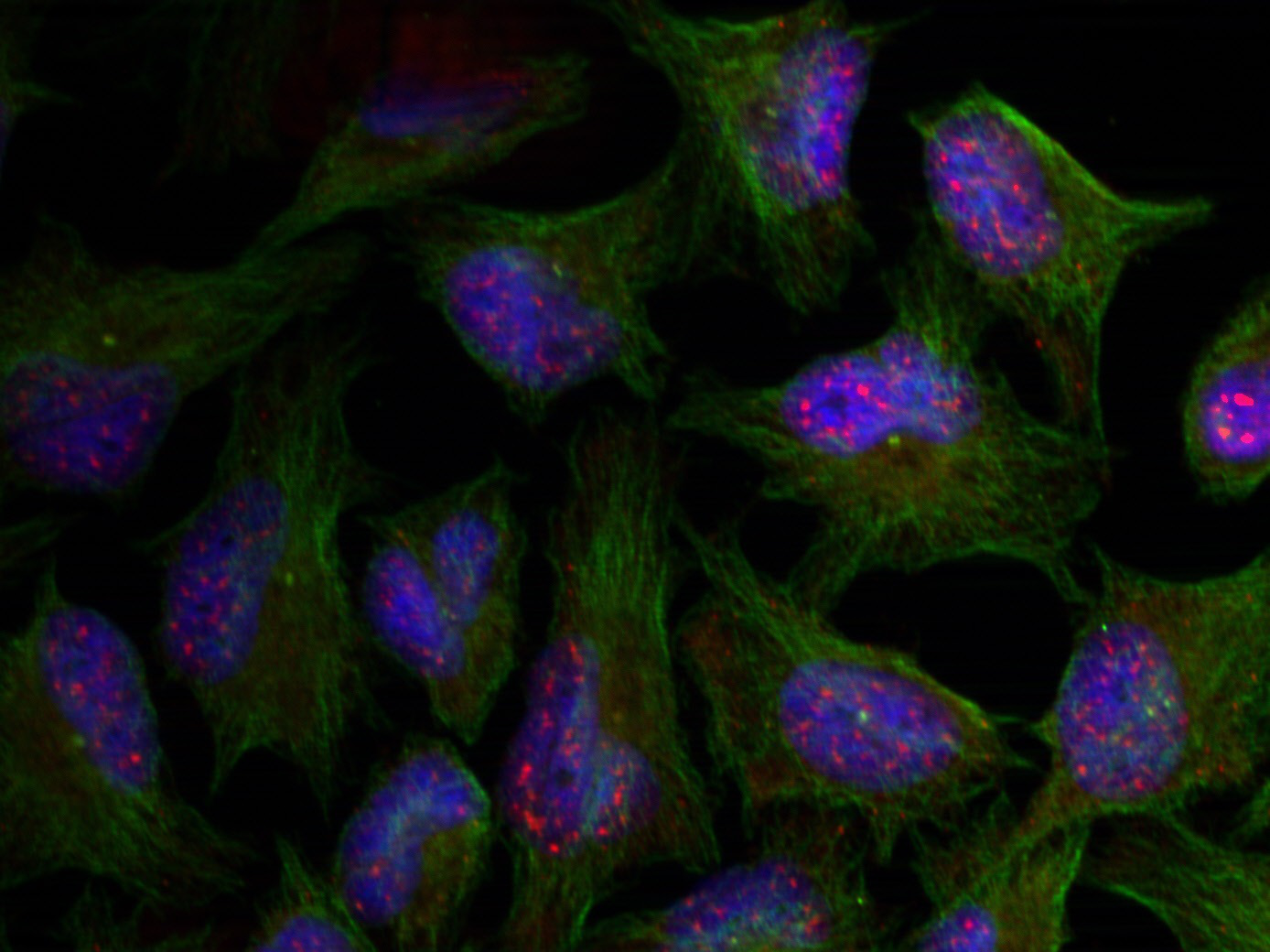
HeLa cell were stained with AF488 conjugated phalloidin (green) and DAPI (blue).
风险提示:丁香通仅作为第三方平台,为商家信息发布提供平台空间。用户咨询产品时请注意保护个人信息及财产安全,合理判断,谨慎选购商品,商家和用户对交易行为负责。对于医疗器械类产品,请先查证核实企业经营资质和医疗器械产品注册证情况。
 文献和实验
文献和实验从一种毒性菇类中分离的剧毒生物碱,它同细胞松弛素的作用相反, 只与聚合的微丝结合, 而不与肌动蛋白单体分子结合。它同聚合的微丝结合后, 抑制了微丝的解体, 因而破坏了微丝的聚合和解聚的动态平衡。
【五分钟讲实验】F-actin 怎么看?一篇讲清鬼笔环肽染色和避坑要点
一、鬼笔环肽是什么? 鬼笔环肽,也常写作 phalloidin,是来源于毒蘑菇的一类环状多肽。 它在实验室里最常见的身份不是“毒素”,而是 F-actin 染色探针。科研人员把它连接上 FITC、TRITC、Alexa Fluor 等荧光基团后,就可以用来观察细胞内聚合态肌动蛋白微丝。 · 它主要识别F-actin,也就是聚合成丝状结构的肌动蛋白,而不是游离的 G-actin 单体。 · 它与 F-actin 结合后能让微丝结构在荧光显微镜或共聚焦显微镜下变得清楚。 · 它通常用于固定
微丝的显示方法步骤:1. 用PBS液漂洗盖片培养的原代细胞3次,每次30s; 2. 用2%的甲醛/PBS液固定原代细胞3min; 3. 用0.5%的三硝基甲苯/PBS处理3次,每次10min; 4. PBS漂洗3次; 5. 用罗丹明(rhodamine)标记的鬼笔环肽(phalloidin)(1:10)室温中反应15min; 6. PBS漂洗3次; 7. 60%甘油+荧光防淬剂封片; 8. 荧光显微镜观察; 微管的显示方法:
 技术资料
技术资料暂无技术资料 索取技术资料

