


- ¥1480
- EASYBIO
- BE3012-100
- 2026年03月11日

企业认证
相关产品推荐更多 >

万千商家帮你免费找货
0 人在求购买到急需产品
- 详细信息
- 文献和实验
- 技术资料
- 形态:
液体
- 保存条件:
-20℃
- 克隆性:
多克隆
- 抗体英文名:
Histone H3 (tri methyl K79) Mouse Monoclonal Antibody
- 规格:
支

Histone H3 is one of the five main histone proteins involved in the structure of chromatin in eukaryotic cells. Core component of nucleosome. Nucleosomes wrap and compact DNA into chromatin, limiting DNA accessibility to the cellular machineries which require DNA as a template. Histones thereby play a central role in transcription regulation, DNA repair, DNA replication and chromosomal stability.
Product Name:Histone H3 (tri methyl K79) Mouse Monoclonal Antibody(4G4)
Clone Number: 4G4
Isotype: IgG1
Storage Buffer :PBS, pH 7.4, containing 0.02% sodium azideas Preservative and 50% Glycerol
Storage instructions:-20°C. Do not aliquot the antibody
Recommended dilutions: WB: 1:500-2,000 IP: 1:200
Optimal dilutions should be determined by the end user.
Specificity: Antibody can detects endogenous Histone H3 (tri methyl K79) protein.
Alternative Names:H3 histone antibody, HIST1H3A antibody, Histone cluster 1, H3a antibody
Molecular Weight:15KD
Form: Liquid
Reactivity:H, R, M
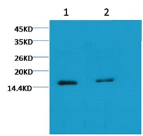
Western blot analysis of Hela with Histone H3 (tri methyl K79) Mouse mAb(4G4) diluted at
1)1:2,000
2)1:5,000
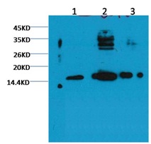
Western blot analysis of 1) Hela, 2) Rat Testis tissue, 3) Raw264.7 with Histone H3 (tri methyl K79) (4G4) Mouse MAb diluted at 1:2000.
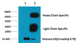
1、Input: Hela Cell Lysate
2、IP product: IP dilute 1:200
Western blot analysis: primary antibody : BE30121:1,000
Secondary antibody: Goat anti-Mouse IgG(H+L) (BE0102) 1:10,000
风险提示:丁香通仅作为第三方平台,为商家信息发布提供平台空间。用户咨询产品时请注意保护个人信息及财产安全,合理判断,谨慎选购商品,商家和用户对交易行为负责。对于医疗器械类产品,请先查证核实企业经营资质和医疗器械产品注册证情况。
 文献和实验
文献和实验Monoclonal Antibody Production
and screening of the hybridomas: 1. Bleeding Mice (1) Place the mouse in a mouse restrainer. (2) Sterilize the tail with 70% ethanol. (3) With a razor blade,nip off the last 2 mm of the tip of the tail. (4) Using a milking motion,pull blood down and let
Detection of Histone H3 Phosphorylation in Cultured Cells and Tissue Sections by Immunostaining
. The protocol described here allows the detection of phosphorylated histones in tissue-cultured cells and tissue sections by fluorescent or bright-field immunostaining analysis. Here we used a serine 10 specific P-histone H3 antibody to determine
FACS-Based Detection of Phosphorylated Histone H3 for the Quantitation of Mitotic Cells
scanner (FACS) is described, based on the presence of an intranuclear antigen present only in mitotic cells, detected using a specific, commercially available antibody. Cell staining and FACS analysis can be done in a single day, making this a rapid
 技术资料
技术资料暂无技术资料 索取技术资料

