

- $132
- ScienCell
- 1201
- 美国
- 2025年07月22日


相关产品推荐更多 >

万千商家帮你免费找货
0 人在求购买到急需产品
- 详细信息
- 文献和实验
- 技术资料
- 库存:
充足
- 英文名:
Pericyte Medium
- 规格:
500ml
产品简介
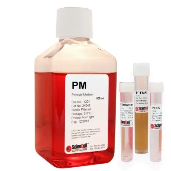
ScienCell® 周细胞培养基(Pericyte Medium,PM)是一款专为人类正常周细胞(Pericytes)体外培养而设计的完整培养系统。结合特定生长添加剂(PGS)和胎牛血清(FBS)使用,可在标准 CO₂ 培养条件下,为周细胞提供理想的生长环境。
产品特色
-
专属配方:精准配比氨基酸、维生素、无机盐、激素、微量元素及生长因子
-
低血清含量(2%):减少干扰因素,促进细胞特异性生长
-
双缓冲系统:HEPES + 碳酸氢盐缓冲,pH 稳定在 7.4(5% CO₂ 培养条件下)
-
无菌即用:所有组分均为液体形式,操作便捷,确保实验重复性
-
专为原代周细胞优化:提高细胞贴壁性与增殖率,适用于形态学、功能及分子实验研究
产品组成
Pericyte Medium(#1201)包含以下组分:
-
基础培养基(500 ml)
-
胎牛血清 FBS(10 ml,货号 #0010)
-
周细胞生长添加剂 PGS(5 ml,货号 #1252)
-
青霉素/链霉素抗生素溶液 P/S(5 ml,货号 #0503)
FBS、PGS 和 P/S 并未预混于基础培养基中,需使用前手动加入,方可组成完整的 Pericyte Medium。
如需询价,请联系我们为您对接中国地区官方供应商。
风险提示:丁香通仅作为第三方平台,为商家信息发布提供平台空间。用户咨询产品时请注意保护个人信息及财产安全,合理判断,谨慎选购商品,商家和用户对交易行为负责。对于医疗器械类产品,请先查证核实企业经营资质和医疗器械产品注册证情况。
 文献和实验
文献和实验-
Md. Mydul Islam, Ignas Gaska, Oluwamayokun Oshinowo, Adiya Otumala, Shashank Shekhar, Nicholas Au Yong, David R. Myers; Single-pericyte nanomechanics measured by contraction cytometry. APL Bioeng. 1 September 2024; 8 (3): 036109.Pericytes line the microvasculature throughout the body and play a key role in regulating blood flow by constricting and dilating vessels. However, the biophysical mechanisms through which pericytes transduce microenvironmental chemical and mechanical cues to mediate vessel diameter, thereby impacting oxygen and nutrient delivery, remain largely unknown. This knowledge gap is clinically relevant as numerous diseases are associated with the aberrant contraction of pericytes, which are unusually susceptible to injury. Here, we report the development of a high-throughput hydrogel-based pericyte contraction cytometer that quantifies single-cell contraction forces from murine and human pericytes in different microvascular microenvironments and in the presence of competing vasoconstricting and vasodilating stimuli. We further show that murine pericyte survival in hypoxia is mediated by the mechanical microenvironment and that, paradoxically, pre-treating pericytes to reduce contraction increases hypoxic cell death. Moreover, using the contraction cytometer as a drug-screening tool, we found that cofilin-1 could be applied extracellularly to release murine pericytes from hypoxia-induced contractile rigor mortis and, therefore, may represent a novel approach for mitigating the long-lasting decrease in blood flow that occurs after hypoxic injury.2.. Rustenhoven J, Smyth LC, Jansson D, Schweder P, Aalderink M, Scotter EL, Mee EW, Faull RLM, Park TI, Dragunow M. (2018). Modelling physiological and pathological conditions to study pericyte biology in brain function and dysfunction. BMC Neurosci. 19(1):6.Background: Brain pericytes ensheathe the endothelium and contribute to formation and maintenance of the blood-brain-barrier. Additionally, pericytes are involved in several aspects of the CNS immune response including scarring, adhesion molecule expression, chemokine secretion, and phagocytosis. In vitro cultures are routinely used to investigate these functions of brain pericytes, however, these are highly plastic cells and can display differing phenotypes and functional responses depending on their culture conditions. Here we sought to investigate how two commonly used culture media, high serum containing DMEM/F12 and low serum containing Pericyte Medium (ScienCell), altered the phenotype of human brain pericytes and neuroinflammatory responses. Methods: Pericytes were isolated from adult human brain biopsy tissue and cultured in DMEM/F12 (D-pericytes) or Pericyte Medium (P-pericytes). Immunocytochemistry, qRT-PCR, and EdU incorporation were used to determine how this altered their basal phenotype, including the expression of pericyte markers, proliferation, and cell morphology. To determine whether culture media altered the inflammatory response in human brain pericytes, immunocytochemistry, qRT-PCR, cytometric bead arrays, and flow cytometry were used to investigate transcription factor induction, chemokine secretion, adhesion molecule expression, migration, phagocytosis, and response to inflammatory-related growth factors. Results: P-pericytes displayed elevated proliferation and a distinct bipolar morphology compared to D-pericytes. Additionally, P-pericytes displayed lower expression of pericyte-associated markers NG2, PDGFRβ, and fibronectin, with notably lower αSMA, CD146, P4H and desmin, and higher Col-IV expression. Nuclear NF-kB translocation in response to IL-1β stimulation was observed in both cultures, however, P-pericytes displayed elevated expression of the transcription factor C/EBPδ, and lower expression of the adhesion molecule ICAM-1. P-pericytes displayed elevated phagocytic and migratory ability. Both cultures responded similarly to stimulation by the growth factors TGFβ1 and PDGF-BB. Conclusions: Despite differences in their phenotype and magnitude of response, both P-pericytes and D-pericytes responded similarly to all examined functions, indicating that the neuroinflammatory phenotype of these cells is robust to culture conditions. Keywords: Blood–brain barrier; Growth factor; Inflammation; Migration; Phagocytosis3. Yao, Y., Norris, E.H., C, E.M. & Strickland, S.(2016) 'Laminin regulates PDGFRb+ cell stemness and muscle development' Nat Commun. VOL 7Muscle-resident PDGFRβ+ cells, which include pericytes and PW1+ interstitial cells (PICs), play a dual role in muscular dystrophy. They can either undergo myogenesis to promote muscle regeneration or differentiate into adipocytes and other cells to compromise regeneration. How the differentiation and fate determination of PDGFRβ+ cells are regulated, however, remains unclear. Here, by utilizing a conditional knockout mouse line, we report that PDGFRβ+ cell-derived laminin inhibits their proliferation and adipogenesis, but is indispensable for their myogenesis. In addition, we show that laminin alone is able to partially reverse the muscle dystrophic phenotype in these mice at the molecular, structural and functional levels. Further RNAseq analysis reveals that laminin regulates PDGFRβ+ cell differentiation/fate determination via gpihbp1. These data support a critical role of laminin in the regulation of PDGFRβ+ cell stemness, identify an innovative target for future drug development and may provide an effective treatment for muscular dystrophy.
 技术资料
技术资料需要更多技术资料 索取更多技术资料
资料下载:
1201-GF.pdf 附 (下载 0 次)
1201.pdf 附 (下载 0 次)
1201-NG.pdf 附 (下载 0 次)
1201-b-prf.pdf 附 (下载 0 次)
1201-b.pdf 附 (下载 0 次)
请 [登录] 后再下载!
