


- ¥3500 - 4500
- 欣润生物(NEWGAINBIO)
- 江苏无锡
- IH1017
- 2026年09月27日

企业认证
相关产品推荐更多 >



万千商家帮你免费找货
0 人在求购买到急需产品
相关阅读
源叶生物 十万种现货试剂速发,品质稳、品类全,实验不再等!Takara 秋促来袭,提取+反转录+定量套装仅 1200 元abinScience 体内级抗体 全年锁价 低至 ¥1200阿拉丁 22万+ 科研级产品现货速发,品质严控、科研无忧- 详细信息
- 文献和实验
- 技术资料
- 英文名:
Immortalized Human Umbilical Artery Endothelial Cells
- 库存:
100万
- 供应商:
欣润生物
- 肿瘤类型:
否
- 细胞类型:
永生化
- ATCC Number:
无
- 品系:
人源
- 组织来源:
脐动脉
- 相关疾病:
无
- 物种来源:
人源
- 免疫类型:
不详
- 细胞形态:
梭形
- 是否是肿瘤细胞:
否
- 器官来源:
脐动脉
- 运输方式:
常温
- 年限:
/
- 生长状态:
贴壁生长
- 规格:
T25方瓶
永生化人脐动脉内皮细胞简介:
产品描述:脐带是胎儿和胎盘之间的连系结构。形状如绳索,表面光滑透明,内含结缔组织和一支脐静脉,一对脐动脉。在子宫中,子宫动脉在胎盘的母体部分出的毛细血管,与胎盘的子体部胎儿毛细血管靠近,在此处母体和胎儿的血液间进行CO2和O2,代谢产物即代谢废物和营养物质的交换。脐动脉将胎儿产生的废物运送至胎盘,脐静脉将O2和营养物质从胎盘运送给胎儿。
产品货号:IH1017
产品类型: 原代细胞建立的永生化
传代能力: 30代左右
产品形态: 梭形
培养基:永生化人脐动脉内皮细胞专用完全培养基,产品编号:IH1017-5
支原体:呈阴性
产品培养条件:37℃,5%CO2
发货方式:常温T25方瓶运输
货期:1周左右货期
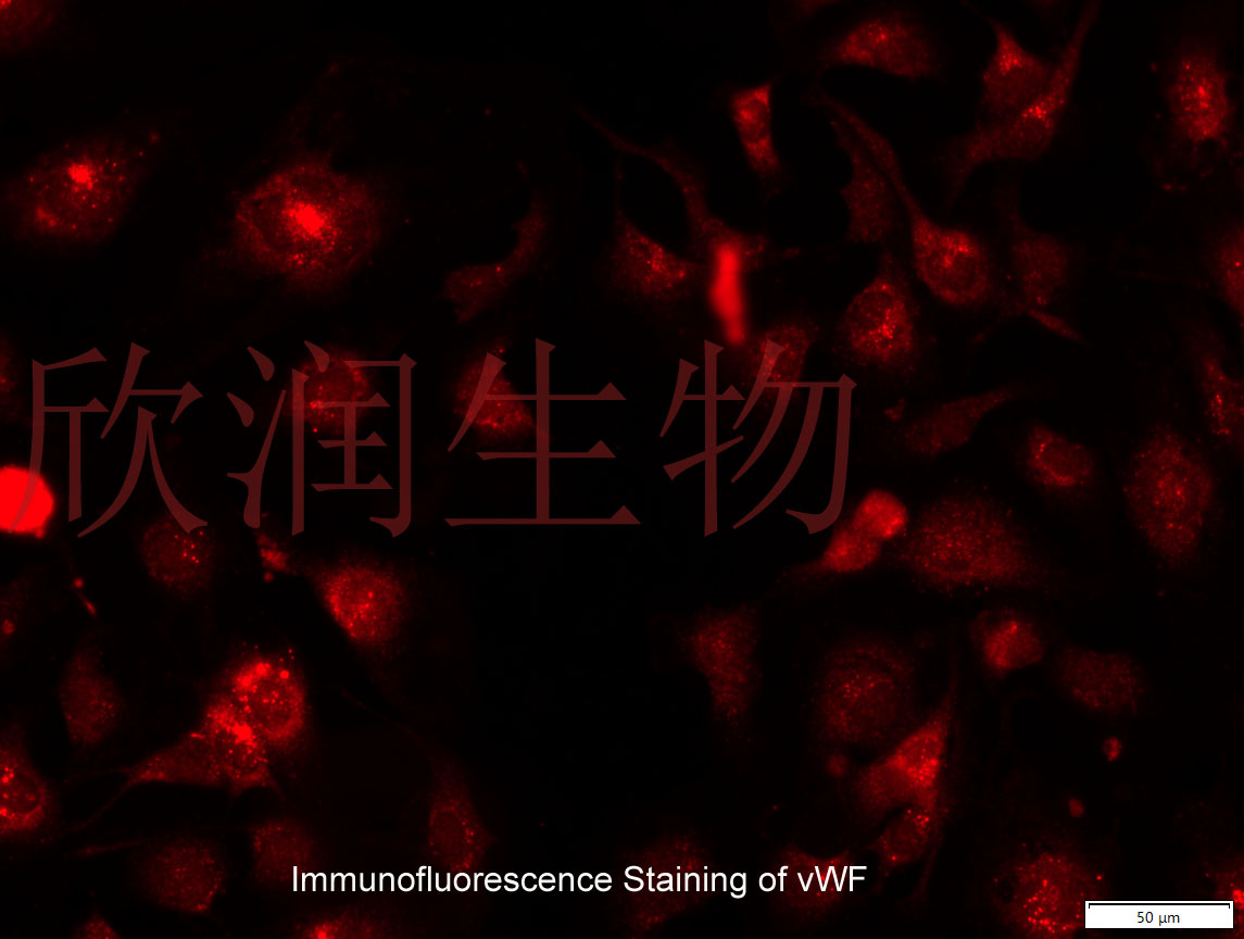
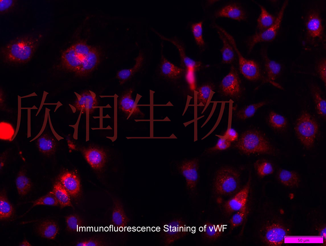
vWF抗体免疫荧光染色鉴定




Phospholipase Cε Modulates Rap1 Activity and the Endothelial BarrierPhospholipase Cε Regulation of Endothelial Rap1
The phosphoinositide-specific phospholipase C, PLCε, is a unique signaling protein with known roles in regulating cardiac myocyte growth, astrocyte inflammatory signaling, and tumor formation. PLCε is also expressed in endothelial cells, however its role in endothelial regulation is not fully established. We show that endothelial cells of multiple origins, including human pulmonary artery (HPAEC), human umbilical vein (HUVEC), and immortalized brain microvascular (hCMEC/D3) endothelial cells, express PLCε. Knockdown of PLCε in arterial endothelial monolayers decreased the effectiveness of the endothelial barrier. Concomitantly, RhoA activity and stress fiber formation were increased. PLCε-deficient arterial endothelial cells also exhibited decreased Rap1-GTP levels, which could be restored by activation of the Rap1 GEF, Epac, to rescue the increase in monolayer leak. Reintroduction of PLCε rescued monolayer leak with both the CDC25 GEF domain and the lipase domain of PLCε required to fully activate Rap1 and to rescue endothelial barrier function. Finally, we demonstrate that
风险提示:丁香通仅作为第三方平台,为商家信息发布提供平台空间。用户咨询产品时请注意保护个人信息及财产安全,合理判断,谨慎选购商品,商家和用户对交易行为负责。对于医疗器械类产品,请先查证核实企业经营资质和医疗器械产品注册证情况。
 文献和实验
文献和实验Chicken intestinal epithelial cells were obtained from NEWGAINBIO company. Cells were cultured on 37℃, with 5% CO2, in the Ham’s F-12 Nutrient (DMEM/12) that contained the following supplementations: fetal bovine serum (5%), in-sulin (5 µg/mL), transferrin (5 µg/mL), selenium (5 ng/mL), epidermal growth factor (5 ng/mL) and penicillin-streptomycin (100–100 U/mL) for cell culturing (full DMEM/12). Experiments were performed with chicken intestinal epithelial cells and working solutions were prepared with plain DMEM/12 without supplementation. For the investigations, cells were seeded onto 96-well, 24-well or 6-well polystyrene cell culture plates.
Primary hVICs (passage 2) were cultured to 50–60% confluence and infected with pGMLV-SV40T-puro lentivirus (NewgainBio, Wuxi, China) at a multiplicity of infection of 80 supplemented with 5 µg/mL polybrene (Sigma-Aldrich, Buchs, Switzerland).
Tissue was cultured until cells became visible around the tissue, and when the fusion reached 90% (FIGURE 1A) §ask ¦lled with the prepared culturing medium was sent to the company for further immortalisation. Cell immortalisation was done for cell stability and longer-term use. Immortalised cells were cultured with 10% FBS and 1% PS in the DMEM medium. After the cells multiplied and merged, they were routinely passed and grown ( NEWGAINBIO Inc. Wuxi, Jiangsu, China) (FIGURE 1B-C).
Mouse primary cultured renal vascular ECs and VSMCs were obtained from Newgainbio company, which were tested by Factor VIII and α-smooth muscle actin (α-SMA), the marker of ECs and VSMCs. RNeasy Mini Kit was used for RNA extraction, and the above protocols were repeated.
Porcine primary colon epithelial cells (Newgainbio company, Wuxi,China) were cultured in Dulbecco's Modified Eagle's Medium (Solarbio, Beijing, China) containing 10 % fetal bovine serum (BioInd, Kiryat shmona, Lsrael) at 37 ◦C and 5 % CO2 humidity.
glutamine-pyruvate-phenol-red-no-hepes.html DMEM (Dulbecco’s Modified Eagle Medium) 是广泛使用的基础培养基,适于培养多种哺乳动物细胞,包括 Hela、293、Cos-7、PC-12 等细胞系,以及原代成纤维细胞、神经元、神经胶质细胞、人脐带静脉内皮细胞、平滑肌细胞等。 青霉/链霉素双抗溶液 www.medchemexpress.cn/inhibitor-kit/penicillin-streptomycin-(100%C3
阿尔茨海默病(AD)研究・体外篇| 从细胞到类器官,体外模型选型手册
体外模型种类很多——从永生化细胞系到原代神经元,从患者来源iPSC到脑类器官和器官芯片,每一类模型在细胞成熟度、实验通量、人源相关性和操作难度上都有明显差异。 本文梳理阿尔茨海默病(AD)各类体外模型的适用场景、优势与短板,搭配选型对照表,方便大家按需挑选合适模型。 一、体外模型主要分为哪几类? 胞系通量高,但生理相关性有限;原代细胞更接近体内,但批次差异明显;iPSC和脑类器官具有人源背景,却面临成熟度和标准化问题。 二、永生化细胞系:操作方便,适合机制
1,主要表达于血管内皮细胞。hFLT1启动子是目前比较常用的血管内皮细胞特异性启动子。 经验分享 高脂饮食会诱导冠状动脉内皮细胞(CAECs)中信号通路失调,进而导致血管张力调节异常、组织灌注改变,并增加冠状动脉疾病的风险。江南大学无锡医学院的研究团队发表了关于调节冠状动脉血管张力的研究成果。研究人员利用和元构建的携带hFLT1启动子的AAV产品,在野生型小鼠血管内皮细胞中特异性过表达KCa3.1和KCa3.1-AR
 技术资料
技术资料
