


- ¥850 - 5500
- 冠导生物
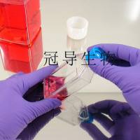
- Caco-2 Cells人结直肠腺癌细胞实验室复苏|STR图谱
- 美国、德国、欧洲等
- 2025年07月15日

1 年金牌会员
企业认证
相关产品推荐更多 >


万千商家帮你免费找货
0 人在求购买到急需产品
- 详细信息
- 文献和实验
- 技术资料
- 品系:
详见细胞说明资料
- 细胞类型:
详见细胞说明资料
- 肿瘤类型:
详见细胞说明资料
- 供应商:
上海冠导生物工程有限公司
- 库存:
≥100瓶
- 生长状态:
详见细胞说明资料
- 年限:
详见细胞说明资料
- 运输方式:
常温运输【复苏细胞】或干冰运输【冻存细胞】
- 器官来源:
详见细胞说明资料
- 是否是肿瘤细胞:
详见细胞说明资料
- 细胞形态:
详见细胞说明资料
- 免疫类型:
详见细胞说明资料
- 物种来源:
详见细胞说明资料
- 相关疾病:
详见细胞说明资料
- 组织来源:
详见细胞说明资料
- 英文名:
Caco-2 Cells人结直肠腺癌细胞实验室复苏|STR图谱
- 规格:
1*10(6)Cellls/瓶
"Caco-2 Cells人结直肠腺癌细胞实验室复苏|STR图谱
BJ Cells人皮肤成纤维细胞保种中心|STR图谱
SW948(DM) Cells人结肠腺癌细胞保种中心|STR图谱
Beta-TC-6 Cells小鼠胰岛素瘤胰岛β细胞保种中心|STR图谱
CMT-U27 Cells犬乳腺癌细胞保种中心|STR图谱
GP293 Cells人胚肾细胞保种中心|STR图谱
HepG2.2.15-LUC Cells人肝细胞保种中心|STR图谱
CTLL-2 Cells小鼠T细胞保种中心|STR图谱
PLC/PRF/5 Cells人肝癌细胞保种中心|STR图谱
物种来源:Caco-2 Cells人结直肠腺癌细胞实验室复苏|STR图谱来源于人源、鼠源等其它物种来源
生长特性:Caco-2 Cells人结直肠腺癌细胞实验室复苏|STR图谱贴壁生长
公司细胞系培养稳定、耐活、复苏好,提供人源、鼠源、兔源、猪源等不同组织如肺、气管、支气管、甲状腺、胰腺、垂体、肾上腺、扁桃体、胸腺、肾、膀胱、输尿管、前列腺、心脏、血管等细胞系。全程提供细胞系组织来源、生长特性、细胞形态、背景资料、培养条件、冻存条件、参考文献、运输方式等产品信息及技术服务。
细胞经过多次传代后,建立了稳定的体外培养模型且可稳定增殖。这些微小的生命单元在培养瓶中生生不息,恰似一支训练有素的军团,为科学家们科研实验提供着源源不断的创新灵感与技术支持。其价值不仅体现在当前广泛的应用中,更在于它为未来生命科学研究提供的可能性。随着技术的进步和对这一细胞系认识的深入,必将在更多领域展现其独特优势。研究人员在使用时应当充分了解其特性,合理设计实验,并注意细胞培养的质量控制,以确保研究结果的可靠性和可重复性。
形态特性:Caco-2 Cells人结直肠腺癌细胞实验室复苏|STR图谱上皮细胞样
换液周期:Caco-2 Cells人结直肠腺癌细胞实验室复苏|STR图谱每周2-3次
产品包装:复苏发货:Caco-2 Cells人结直肠腺癌细胞实验室复苏|STR图谱T25培养瓶(一瓶)或冻存发货:1ml冻存管(两支)
在细胞培养的日常操作中,细胞消化是一个看似简单却极易被误解的关键步骤。许多研究者,尤其是初学者,往往陷入过度消化的误区,认为必须将细胞吹打成完全离散的单细胞悬液才算消化完成。然而,这种认知不仅缺乏科学依据,更可能对细胞造成不可逆的损伤。事实上,绝大部分贴壁细胞的消化只需采用""胰酶润洗法""即可高效完成,即用胰酶润洗一遍后吸去,残留的微量胰酶在37℃条件下通常能在1-2分钟内使细胞充分脱离。这种方法的精妙之处在于利用难以精确计量的残留胰酶实现温和消化,既避免了酶解过度,又维持了细胞活性。
ATCC细胞库(American Type Culture Colection),该中心一直致力于细胞分类、鉴定和保藏工作。ATCC是全球最大的生物资源保藏中心,ATCC通过行业标准产品、服务和创新解决方案支持全球学术、政府、生物技术、制药、食品、农业和工业领域的科学进步。ATCC提供的服务和定制解决方案包括细胞和微生物培养、鉴定、生物衍生物的开发和生产、性能测试和生物资源保藏服务。美国国家标准协会(ANSI)认可了ATCC标准开发组织,并制定了标准协议,以确保生物材料的可靠性和可重复性。ATCC的使命是为了获取、鉴定、保存、开发、标准化和分发生物资源和生物信息,以提高和应用生物科学知识。
背景信息:Caco-2 Cells人结直肠腺癌细胞实验室复苏|STR图谱细胞株分离自一个原发性结肠癌。当细胞长满时,表现出典型的肠细胞分化的特征。Caco-2细胞表达维生素A结合蛋白I和视黄醇结合蛋白II,角蛋白阳性。
传代比例:1:2-1:4(Caco-2 Cells人结直肠腺癌细胞实验室复苏|STR图谱首次传代建议1:2)
来源说明:Caco-2 Cells人结直肠腺癌细胞实验室复苏|STR图谱主要来源ATCC、ECACC、DSMZ、RCB等细胞库
营养供给是细胞的生命基石。就像新生儿需要母乳或配方奶粉提供全面营养,初代细胞或刚刚复苏的冻存细胞对营养环境有着近乎苛刻的要求。科研人员会为这些""细胞婴儿""精心配制含有10-15%胎牛血清的基础培养基,这相当于为细胞准备的高级""配方奶粉""。血清中富含的生长因子、激素和附着因子,就像母乳中的免疫球蛋白和益生菌,能为细胞提供最贴心的保护。而对于某些娇贵的干细胞或免疫细胞,还需要额外添加EGF、bFGF等""营养强化剂"",这些细胞因子相当于婴幼儿的DHA和维生素补充剂,能显著提高细胞的贴壁率和增殖活性。实验室数据表明,在复苏后24小时内添加适量血清的细胞,其存活率可比常规培养提高30%以上。
血清对温度极为敏感,必须严格控制在–20℃至–70℃的环境中保存。这一温度范围能够有效维持血清中各种生物活性成分的稳定性,包括生长因子、激素、蛋白质等。若因特殊情况需要将血清暂时存放于4℃冰箱,务必注意时间不得超过一个月,否则血清中的有效成分会逐渐降解,影响细胞培养效果。对于使用频率较低的实验室,建议将血清保存在–70℃超低温冰箱中,以最大限度延长其保质期。值得注意的是,即使在–20℃条件下,血清也不宜存放过久,通常建议在一年内使用完毕。
HKb20 Cells人肾上皮细胞保种中心|STR图谱
SK-MEL-1 Cells人皮肤黑色素瘤细胞保种中心|STR图谱
B16-RFP Cells小鼠黑色素瘤细胞保种中心|STR图谱
5637-Luc Cells人膀胱癌细胞保种中心|STR图谱
Sf21 Cells;背景说明:详见相关文献介绍;传代方法:1:2-1:3传代;每周换液2-3次。;生长特性:贴壁或悬浮,详见产品说明书部分;形态特性:详见产品说明书;相关产品有:U-251_MG细胞、NHRF细胞、HT-3细胞
Hs-343-T Cells;背景说明:详见相关文献介绍;传代方法:1:2—1:3传代;每周换液2-3次。;生长特性:贴壁生长;形态特性:成纤维细胞;相关产品有:FL83B细胞、RCK-8细胞、HCT FET细胞
CCK81 Cells;背景说明:详见相关文献介绍;传代方法:1:2-1:3传代;每周换液2-3次。;生长特性:贴壁或悬浮,详见产品说明书部分;形态特性:详见产品说明书;相关产品有:HLE-B3细胞、IOSE29细胞、NCI H23细胞
GM11889 Cells(提供STR鉴定图谱)
HAP1 DIAPH1 (-) 2 Cells(提供STR鉴定图谱)
B16-Luc Cells小鼠黑色素瘤细胞保种中心|STR图谱
U-87MG-RFP Cells人脑星形胶质母细胞瘤细胞保种中心|STR图谱
lewis-luc Cells小鼠肺癌细胞保种中心|STR图谱
OCM-1A-RFP Cells人眼脉络黑色素瘤细胞保种中心|STR图谱
CCD-841CoN Cells人正常结肠上皮细胞保种中心|STR图谱
Anglne Cells人卵巢癌细胞保种中心|STR图谱
WSU-DLCL(2) Cells;背景说明:弥漫大B淋巴瘤;男性;传代方法:1:2-1:3传代;每周换液2-3次。;生长特性:悬浮;形态特性:详见产品说明书;相关产品有:MDCK细胞、V 79-4细胞、HCV 29细胞
H-4 Cells;背景说明:H4细胞系建系于1973年。它衍生于一个患神经胶质瘤的37岁病人的脑组织。该细胞的致瘤特性己经被屏蔽,细胞接种动物一般不产生肿瘤结节。该细胞具有修复MNNG损伤5型腺病毒的能力。;传代方法:1:3传代,每周换液2-3次。;生长特性:贴壁生长;形态特性:上皮样;相关产品有:MV522细胞、EOC20细胞、RBL 1细胞
TE-3A Cells;背景说明:详见相关文献介绍;传代方法:1:2-1:3传代;每周换液2-3次。;生长特性:贴壁或悬浮,详见产品说明书部分;形态特性:详见产品说明书;相关产品有:COLO 741细胞、GM06141B细胞、SuperTube细胞
Leukemia L1210 Cells;背景说明:该细胞源于用0.2%甲基胆蒽(溶解)涂抹雌性小鼠的皮肤诱发的肿瘤,鼠痘病毒阴性。;传代方法:1:2传代;生长特性:悬浮生长;形态特性:淋巴母细胞样;相关产品有:Normal Rat, August 3, 1983细胞、NCI.H226细胞、HCGC细胞
PTK2 Cells;背景说明:详见相关文献介绍;传代方法:1:2-1:3传代;每周换液2-3次。;生长特性:贴壁或悬浮,详见产品说明书部分;形态特性:详见产品说明书;相关产品有:JIII细胞、OVCAR3细胞、MDA-MB-435 S细胞
FDC-P1 Cells;背景说明:详见相关文献介绍;传代方法:2-3天换液1次;生长特性:悬浮生长;形态特性:淋巴母细胞;相关产品有:CMT-93细胞、NCTC clone 929细胞、COLO 829细胞
Medical University of Graz-Chordoma 1 Cells;背景说明:骶骨脊索瘤;女性;传代方法:1:2-1:3传代;每周换液2-3次。;生长特性:贴壁;形态特性:详见产品说明书;相关产品有:HEH2细胞、CA46细胞、B95-8细胞
KU-19-19 Cells人膀胱癌细胞保种中心|STR图谱
HEY Cells人卵巢癌细胞保种中心|STR图谱
HGC-27+YFP Cells人胃癌细胞保种中心|STR图谱
MUM2B Cells人侵袭性脉络膜黑色素瘤细胞保种中心|STR图谱
JHH-2 Cells人肝癌细胞保种中心|STR图谱
DU4475 Cells人乳腺上皮细胞保种中心|STR图谱
MEL Cells小鼠红白血病细胞保种中心|STR图谱
HGC27 Cells;背景说明:未分化胃癌,能分泌粘液素。;传代方法:消化3-5分钟,1:2,3天内可长满;生长特性:贴壁生长;形态特性:上皮样;相关产品有:SK BR 03细胞、MDA-134细胞、Hep G2细胞
HHUUKDi007-A Cells(提供STR鉴定图谱)
iOSE-C9 Cells(提供STR鉴定图谱)
Ma-Mel-79 Cells(提供STR鉴定图谱)
ND08030 Cells(提供STR鉴定图谱)
SEWA/TC13 Cells(提供STR鉴定图谱)
NIH/3T3-LUC Cells小鼠胚胎成纤维细胞保种中心|STR图谱
PC-12-GFP Cells大鼠肾上腺嗜铬细胞瘤细胞保种中心|STR图谱
PFIZi010-B Cells(提供STR鉴定图谱)
Ubigene ID8 Kmt2c KO Cells(提供STR鉴定图谱)
YAN3141 Cells(提供STR鉴定图谱)
HG02809 Cells(提供STR鉴定图谱)
HN 4 Cells;背景说明:喉鳞癌;男性;传代方法:1:2-1:3传代;每周换液2-3次。;生长特性:贴壁;形态特性:详见产品说明书;相关产品有:NTERA2细胞、EFM-192A细胞、NCI-H1915细胞
MDBK Cells;背景说明:详见相关文献介绍;传代方法:1:2传代;生长特性:贴壁生长 ;形态特性:详见产品说明书;相关产品有:ECC10细胞、U-hth74细胞、HBZY1细胞
251MG Cells;背景说明:U-251 MG分离至一位患者的胶质母细胞瘤组织。;传代方法:1:2-1:3传代;每周换液2-3次。;生长特性:贴壁;形态特性:成纤维细胞样;相关产品有:Hs-578T细胞、IMCD-3细胞、HN 4细胞
MV 3 Cells;背景说明:黑色素瘤;淋巴结转移;男性;传代方法:1:2-1:3传代;每周换液2-3次。;生长特性:贴壁;形态特性:详见产品说明书;相关产品有:MDAMB361细胞、UCLA SO M21细胞、Stanford University Pediatric T-cell line 1细胞
OVCAR-3 Cells人卵巢癌细胞保种中心|STR图谱
143B Cells人骨肉瘤细胞保种中心|STR图谱
IM95m Cells人胃腺癌细胞保种中心|STR图谱
BT483 Cells人乳腺导管癌细胞保种中心|STR图谱
Panc 03.27 Cells人胰腺癌细胞保种中心|STR图谱
NIH 3T3-RFP Cells小鼠胚胎成纤维细胞保种中心|STR图谱
MC38 Cells;背景说明:详见相关文献介绍;传代方法:1:2传代;生长特性:贴壁生长;形态特性:上皮细胞样;相关产品有:QG56细胞、H-1238细胞、OCM1A细胞
Human fetal lung fibroblast 1 Cells;背景说明:详见相关文献介绍;传代方法:消化3-5分钟。1:2。3天内可长满。;生长特性:贴壁生长;形态特性:成纤维细胞样;相关产品有:OCI AML3细胞、LC-1 sq细胞、WM239细胞
S37 Cells;背景说明:肉瘤 Cells;传代方法:1:2-1:3传代;每周换液2-3次。;生长特性:贴壁;形态特性:详见产品说明书;相关产品有:HBE135-E6E7细胞、Jurkat, Clone E6-1细胞、NCIH716细胞
S37 Cells;背景说明:肉瘤 Cells;传代方法:1:2-1:3传代;每周换液2-3次。;生长特性:贴壁;形态特性:详见产品说明书;相关产品有:HBE135-E6E7细胞、Jurkat, Clone E6-1细胞、NCIH716细胞
S.B. Cells;背景说明:急性T淋巴细胞白血病;男性;传代方法:1:2-1:3传代;每周换液2-3次。;生长特性:悬浮;形态特性:详见产品说明书;相关产品有:BEL7402细胞、HEK-293-EBNA细胞、DU 145细胞
SNU-620 Cells;背景说明:详见相关文献介绍;传代方法:1:2-1:3传代;每周换液2-3次。;生长特性:贴壁或悬浮,详见产品说明书部分;形态特性:详见产品说明书;相关产品有:DoHH2细胞、NPC-TW01细胞、NCIH1435细胞
ACHN Cells人肾细胞腺癌细胞保种中心|STR图谱
5637-Luc Cells人膀胱癌细胞保种中心|STR图谱
MDS-L Cells人骨髓增生异常综合征细胞保种中心|STR图谱
HCT 15 Cells人结直肠腺癌细胞保种中心|STR图谱
Jurkat, Clone E6-1-Luc Cells人急性T淋巴细胞白血病细胞保种中心|STR图谱
FTC-133 Cells人滤泡状甲状腺癌细胞保种中心|STR图谱
GP5d Cells;背景说明:结肠癌;女性;传代方法:1:2-1:3传代;每周换液2-3次。;生长特性:贴壁;形态特性:详见产品说明书;相关产品有:SKES-1细胞、OCI-AML2细胞、Vero细胞
KM-12 Cells;背景说明:详见相关文献介绍;传代方法:1:2传代;生长特性:贴壁生长;形态特性:上皮样;相关产品有:T8细胞、MZCRC1细胞、U-251 MG细胞
A375mel Cells;背景说明:A375源自一位54岁女性,是Giard DJ等人建立的一系列细胞株中的一株。该细胞可在免疫抑制小鼠上成瘤,在琼脂上形成克隆。;传代方法:1:2-1:3传代;每周换液2-3次。;生长特性:贴壁;形态特性:上皮细胞样;相关产品有:SKLMS-1细胞、SGC-7901细胞、JHH2细胞
KRC/Y Cells;背景说明:肾透明细胞癌;女性;传代方法:1:2-1:3传代;每周换液2-3次。;生长特性:贴壁;形态特性:详见产品说明书;相关产品有:VMM5细胞、GT1-1细胞、P31/FUJ细胞
S-16 Cells;背景说明:Schwann细胞;自发永生;传代方法:1:2-1:3传代;每周换液2-3次。;生长特性:贴壁;形态特性:详见产品说明书;相关产品有:Hs578Bst细胞、Vero 76 clone E-6细胞、QGY 7701细胞
SVEC4-10 Cells小鼠淋巴结内皮细胞保种中心|STR图谱
NCI-H295R Cells人肾上腺皮质腺癌细胞保种中心|STR图谱
HUVEC+RFP Cells人脐静脉内皮细胞保种中心|STR图谱
NCI-H1650 Cells人非小细胞肺癌细胞保种中心|STR图谱
DT40 Cells鸡淋巴瘤细胞保种中心|STR图谱
NU-DUL-1 Cells人弥漫性组织细胞性淋巴瘤细胞保种中心|STR图谱
MAC-T Cells牛乳腺上皮细胞保种中心|STR图谱
Fao Cells;背景说明:详见相关文献介绍;传代方法:1:2-1:3传代;每周换液2-3次。;生长特性:贴壁或悬浮,详见产品说明书部分;形态特性:上皮细胞;相关产品有:RPMI #8226细胞、Keio University-19-19细胞、BEL 7402细胞
CL1.0 Cells;背景说明:肺癌;男性;传代方法:1:2-1:3传代;每周换液2-3次。;生长特性:贴壁;形态特性:详见产品说明书;相关产品有:COR-L 105细胞、M14-MEL细胞、MMAc-Serum Free细胞
WI-38 Cells;背景说明:LeonardHayflick建系;有限传代细胞系;寿命为50±10代(倍增时间24h);来自妊娠3个月的正常胚胎肺组织。该细胞系是第一个用于人制备的人二倍体细胞;培养基中添加TNFα可以加快细胞生长。;传代方法:1:2-1:4传代;2-3天换液1次;生长特性:贴壁生长;形态特性:成纤维细胞样;相关产品有:RD细胞、LMTK-细胞、OCI-AML3细胞
PANC403 Cells;背景说明:详见相关文献介绍;传代方法:1:2传代;生长特性:贴壁生长;形态特性:上皮样;相关产品有:MFM223细胞、Hs294T细胞、H-929细胞
PC-3M-IE8 Cells;背景说明:前列腺癌;男性;传代方法:1:2-1:3传代;每周换液2-3次。;生长特性:贴壁;形态特性:详见产品说明书;相关产品有:MDA-MB-231-GFP细胞、M059J细胞、CCK-81细胞
Hs 729T Cells;背景说明:详见相关文献介绍;传代方法:1:2传代;每周换液2-3次。;生长特性:贴壁生长;形态特性:成纤维细胞;相关产品有:MDA175细胞、3T3-F442A细胞、SW1463细胞
WEHI231 Cells;背景说明:详见相关文献介绍;传代方法:1:2-1:3传代;每周换液2-3次。;生长特性:贴壁或悬浮,详见产品说明书部分;形态特性:详见产品说明书;相关产品有:LICR-HN6细胞、McA-RH 7777细胞、VMRC-RCZ细胞
SUM-149 Cells;背景说明:乳腺癌;女性;传代方法:1:2-1:3传代;每周换液2-3次。;生长特性:贴壁;形态特性:详见产品说明书;相关产品有:RAMOS2G64C10细胞、HMEC-1细胞、KYSE-150细胞
" "PubMed=7459858
Rousset M., Zweibaum A., Fogh J.
Presence of glycogen and growth-related variations in 58 cultured human tumor cell lines of various tissue origins.
Cancer Res. 41:1165-1170(1981)
PubMed=3518877; DOI=10.3109/07357908609038260
Fogh J.
Human tumor lines for cancer research.
Cancer Invest. 4:157-184(1986)
PubMed=3349466
Chantret I., Barbat A., Dussaulx E., Brattain M.G., Zweibaum A.
Epithelial polarity, villin expression, and enterocytic differentiation of cultured human colon carcinoma cells: a survey of twenty cell lines.
Cancer Res. 48:1936-1942(1988)
PubMed=2914637; DOI=10.1016/0016-5085(89)90897-4
Hidalgo I.J., Raub T.J., Borchardt R.T.
Characterization of the human colon carcinoma cell line (Caco-2) as a model system for intestinal epithelial permeability.
Gastroenterology 96:736-749(1989)
PubMed=7764660; DOI=10.1007/BF00749813
Hashimoto K., Shimizu M.
Epithelial properties of human intestinal Caco-2 cells cultured in a serum-free medium.
Cytotechnology 13:175-184(1993)
PubMed=8253353; DOI=10.1016/0016-5085(93)91076-t
Chastre E., Empereur S., Di Gioia Y., El Mahdani N., Mareel M.M., Vleminckx K.L., Van Roy F.M., Bex V., Emami S., Spandidos D.A., Gespach C.
Neoplastic progression of human and rat intestinal cell lines after transfer of the ras and polyoma middle T oncogenes.
Gastroenterology 105:1776-1789(1993)
PubMed=8508948; DOI=10.1111/j.1432-0436.1993.tb00645.x
de Bruine A.P., de Vries J.E., Dinjens W.N.M., Moerkerk P.T.M., van der Linden E.P.M., Pijls M.M.J., ten Kate J., Bosman F.T.
Human Caco-2 cells transfected with c-Ha-Ras as a model for endocrine differentiation in the large intestine.
Differentiation 53:51-60(1993)
PubMed=9294210; DOI=10.1073/pnas.94.19.10330; PMCID=PMC23362
Ilyas M., Tomlinson I.P.M., Rowan A.J., Pignatelli M., Bodmer W.F.
Beta-catenin mutations in cell lines established from human colorectal cancers.
Proc. Natl. Acad. Sci. U.S.A. 94:10330-10334(1997)
PubMed=10092214; DOI=10.1002/(SICI)1097-4652(199812)177:4<618::AID-JCP12>3.0.CO;2-R
Levy P., Robin H., Kornprobst M., Capeau J., Cherqui G.
Enterocytic differentiation of the human Caco-2 cell line correlates with alterations in integrin signaling.
J. Cell. Physiol. 177:618-627(1998)
PubMed=10612807; DOI=10.1002/(SICI)1098-2264(200002)27:2<183::AID-GCC10>3.0.CO;2-P; PMCID=PMC4721570
Ghadimi B.M., Sackett D.L., Difilippantonio M.J., Schrock E., Neumann T., Jauho A., Auer G., Ried T.
Centrosome amplification and instability occurs exclusively in aneuploid, but not in diploid colorectal cancer cell lines, and correlates with numerical chromosomal aberrations.
Genes Chromosomes Cancer 27:183-190(2000)
PubMed=10737795; DOI=10.1073/pnas.97.7.3352; PMCID=PMC16243
Rowan A.J., Lamlum H., Ilyas M., Wheeler J.M.D., Straub J., Papadopoulou A., Bicknell D.C., Bodmer W.F., Tomlinson I.P.M.
APC mutations in sporadic colorectal tumors: a mutational 'hotspot' and interdependence of the 'two hits'.
Proc. Natl. Acad. Sci. U.S.A. 97:3352-3357(2000)
PubMed=11414198; DOI=10.1007/s004320000207
Lahm H., Andre S., Hoeflich A., Fischer J.R., Sordat B., Kaltner H., Wolf E., Gabius H.-J.
Comprehensive galectin fingerprinting in a panel of 61 human tumor cell lines by RT-PCR and its implications for diagnostic and therapeutic procedures.
J. Cancer Res. Clin. Oncol. 127:375-386(2001)
PubMed=11416159; DOI=10.1073/pnas.121616198; PMCID=PMC35459
Masters J.R.W., Thomson J.A., Daly-Burns B., Reid Y.A., Dirks W.G., Packer P., Toji L.H., Ohno T., Tanabe H., Arlett C.F., Kelland L.R., Harrison M., Virmani A.K., Ward T.H., Ayres K.L., Debenham P.G.
Short tandem repeat profiling provides an international reference standard for human cell lines.
Proc. Natl. Acad. Sci. U.S.A. 98:8012-8017(2001)
PubMed=11668190; DOI=10.1177/002215540104901105
Quentmeier H., Osborn M., Reinhardt J., Zaborski M., Drexler H.G.
Immunocytochemical analysis of cell lines derived from solid tumors.
J. Histochem. Cytochem. 49:1369-1378(2001)
PubMed=12584437; DOI=10.1159/000068544
Melcher R., Koehler S., Steinlein C., Schmid M., Mueller C.R., Luehrs H., Menzel T., Scheppach W., Moerk H., Scheurlen M., Koehrle J., Al-Taie O.
Spectral karyotype analysis of colon cancer cell lines of the tumor suppressor and mutator pathway.
Cytogenet. Genome Res. 98:22-28(2002)
PubMed=14599474; DOI=10.1016/S0887-2333(03)00095-X
Ranaldi G., Consalvo R., Sambuy Y., Scarino M.L.
Permeability characteristics of parental and clonal human intestinal Caco-2 cell lines differentiated in serum-supplemented and serum-free media.
Toxicol. In Vitro 17:761-767(2003)
PubMed=15316659; DOI=10.1007/s00018-004-4222-9; PMCID=PMC7079841
Cinatl J. Jr., Hoever G., Morgenstern B., Preiser W., Vogel J.-U., Hofmann W.-K., Bauer G., Michaelis M., Rabenau H.F., Doerr H.-W.
Infection of cultured intestinal epithelial cells with .
Cell. Mol. Life Sci. 61:2100-2112(2004)
PubMed=15731278; DOI=10.1128/JVI.79.6.3846-3850.2005; PMCID=PMC1075706
Mossel E.C., Huang C., Narayanan K., Makino S., Tesh R.B., Peters C.J.
Exogenous ACE2 expression allows refractory cell lines to support replication.
J. Virol. 79:3846-3850(2005)
PubMed=15868485; DOI=10.1007/s10565-005-0085-6
Sambuy Y., De Angelis I., Ranaldi G., Scarino M.L., Stammati A., Zucco F.
The Caco-2 cell line as a model of the intestinal barrier: influence of cell and culture-related factors on Caco-2 cell functional characteristics.
Cell Biol. Toxicol. 21:1-26(2005)
PubMed=16418264; DOI=10.1073/pnas.0510146103; PMCID=PMC1327731
Liu Y., Bodmer W.F.
Analysis of p53 mutations and their expression in 56 colorectal cancer cell lines.
Proc. Natl. Acad. Sci. U.S.A. 103:976-981(2006)
PubMed=16854228; DOI=10.1186/1476-4598-5-29; PMCID=PMC1550420
Bandres Elizalde E.M., Cubedo E., Agirre X., Malumbres R., Zarate R., Ramirez N., Abajo A., Navarro A., Moreno I., Monzo M., Garcia-Foncillas J.
Identification by real-time PCR of 13 mature microRNAs differentially expressed in colorectal cancer and non-tumoral tissues.
Mol. Cancer 5:29.1-29.10(2006)
PubMed=18258742; DOI=10.1073/pnas.0712176105; PMCID=PMC2268141
Emaduddin M., Bicknell D.C., Bodmer W.F., Feller S.M.
Cell growth, global phosphotyrosine elevation, and c-Met phosphorylation through Src family kinases in colorectal cancer cells.
Proc. Natl. Acad. Sci. U.S.A. 105:2358-2362(2008)
PubMed=20570890; DOI=10.1158/0008-5472.CAN-10-0192; PMCID=PMC2943514
Janakiraman M., Vakiani E., Zeng Z.-S., Pratilas C.A., Taylor B.S., Chitale D., Halilovic E., Wilson M., Huberman K., Ricarte Filho J.C.M., Persaud Y., Levine D.A., Fagin J.A., Jhanwar S.C., Mariadason J.M., Lash A., Ladanyi M., Saltz L.B., Heguy A., Paty P.B., Solit D.B.
Genomic and biological characterization of exon 4 KRAS mutations in human cancer.
Cancer Res. 70:5901-5911(2010)
PubMed=20606684; DOI=10.1038/sj.bjc.6605780; PMCID=PMC2920028
Bracht K., Nicholls A.M., Liu Y., Bodmer W.F.
5-fluorouracil response in a large panel of colorectal cancer cell lines is associated with mismatch repair deficiency.
Br. J. Cancer 103:340-346(2010)
PubMed=20831567; DOI=10.1111/j.1582-4934.2010.01170.x; PMCID=PMC3918049
Ma Y.-L., Zhang P., Wang F., Moyer M.P., Yang J.-J., Liu Z.-H., Peng J.-Y., Chen H.-Q., Zhou Y.-K., Liu W.-J., Qin H.-L.
Human embryonic stem cells and metastatic colorectal cancer cells shared the common endogenous human microRNA-26b.
J. Cell. Mol. Med. 15:1941-1954(2011)
PubMed=21607810; DOI=10.1208/s12248-011-9283-8; PMCID=PMC3160153
Borchardt R.T.
Hidalgo, I.J., Raub, T.J., and Borchardt, R.T.: Characterization of the human colon carcinoma cell line (Caco-2) as a model system for intestinal epithelial permeability, Gastroenterology, 96, 736-749, 1989 -- the backstory.
AAPS J. 13:323-327(2011)
PubMed=23272949; DOI=10.1186/1755-8794-5-66; PMCID=PMC3543849
Schlicker A., Beran G., Chresta C.M., McWalter G., Pritchard A., Weston S., Runswick S., Davenport S., Heathcote K., Castro D.A., Orphanides G., French T., Wessels L.F.A.
Subtypes of primary colorectal tumors correlate with response to targeted treatment in colorectal cell lines.
BMC Med. Genomics 5:66.1-66.15(2012)
PubMed=23932154; DOI=10.1016/j.radonc.2013.06.032
Salendo J., Spitzner M., Kramer F., Zhang X., Jo P., Wolff H.A., Kitz J., Kaulfuss S., Beissbarth T., belstein M., Ghadimi M., Grade M., Gaedcke J.
Identification of a microRNA expression signature for chemoradiosensitivity of colorectal cancer cells, involving miRNAs-320a, -224, -132 and let7g.
Radiother. Oncol. 108:451-457(2013)
PubMed=24042735; DOI=10.1038/oncsis.2013.35; PMCID=PMC3816225
Ahmed D., Eide P.W., Eilertsen I.A., Danielsen S.A., Eknaes M., Hektoen M., Lind G.E., Lothe R.A.
Epigenetic and genetic features of 24 colon cancer cell lines.
Oncogenesis 2:e71.1-e71.8(2013)
PubMed=24755471; DOI=10.1158/0008-5472.CAN-14-0013
Mouradov D., Sloggett C., Jorissen R.N., Love C.G., Li S., Burgess A.W., Arango D., Strausberg R.L., Buchanan D., Wormald S., O'Connor L., Wilding J.L., Bicknell D.C., Tomlinson I.P.M., Bodmer W.F., Mariadason J.M., Sieber O.M.
Colorectal cancer cell lines are representative models of the main molecular subtypes of primary cancer.
Cancer Res. 74:3238-3247(2014)
PubMed=25960936; DOI=10.4161/21624011.2014.954893; PMCID=PMC4355981
Boegel S., Lower M., Bukur T., Sahin U., Castle J.C.
A catalog of HLA type, HLA expression, and neo-epitope candidates in human cancer cell lines.
OncoImmunology 3:e954893.1-e954893.12(2014)
PubMed=25485619; DOI=10.1038/nbt.3080
Klijn C., Durinck S., Stawiski E.W., Haverty P.M., Jiang Z.-S., Liu H.-B., Degenhardt J., Mayba O., Gnad F., Liu J.-F., Pau G., Reeder J., Cao Y., Mukhyala K., Selvaraj S.K., Yu M.-M., Zynda G.J., Brauer M.J., Wu T.D., Gentleman R.C., Manning G., Yauch R.L., Bourgon R., Stokoe D., Modrusan Z., Neve R.M., de Sauvage F.J., Settleman J., Seshagiri S., Zhang Z.-M.
A comprehensive transcriptional portrait of human cancer cell lines.
Nat. Biotechnol. 33:306-312(2015)
PubMed=25841592; DOI=10.1016/j.jprot.2015.03.019
Piersma S.R., Knol J.C., de Reus I., Labots M., Sampadi B.K., Pham T.V., Ishihama Y., Verheul H.M.W., Jimenez C.R.
Feasibility of label-free phosphoproteomics and application to base-line signaling of colorectal cancer cell lines.
J. Proteomics 127:247-258(2015)
PubMed=25877200; DOI=10.1038/nature14397
Yu M., Selvaraj S.K., Liang-Chu M.M.Y., Aghajani S., Busse M., Yuan J., Lee G., Peale F.V., Klijn C., Bourgon R., Kaminker J.S., Neve R.M.
A resource for cell line authentication, annotation and quality control.
Nature 520:307-311(2015)
PubMed=25926053; DOI=10.1038/ncomms8002
Medico E., Russo M., Picco G., Cancelliere C., Valtorta E., Corti G., Buscarino M., Isella C., Lamba S., Martinoglio B., Veronese S., Siena S., Sartore-Bianchi A., Beccuti M., Mottolese M., Linnebacher M., Cordero F., Di Nicolantonio F., Bardelli A.
The molecular landscape of colorectal cancer cell lines unveils clinically actionable kinase targets.
Nat. Commun. 6:7002.1-7002.10(2015)
PubMed=25944804; DOI=10.1158/1078-0432.CCR-14-2457
Bazzocco S., Dopeso H., Carton-Garcia F., Macaya I., Andretta E., Chionh F., Rodrigues P., Garrido M., Alazzouzi H., Nieto R., Sanchez A., Schwartz S. Jr., Bilic J., Mariadason J.M., Arango D.
Highly expressed genes in rapidly proliferating tumor cells as new targets for colorectal cancer treatment.
Clin. Cancer Res. 21:3695-3704(2015)
PubMed=26589293; DOI=10.1186/s13073-015-0240-5; PMCID=PMC4653878
Scholtalbers J., Boegel S., Bukur T., Byl M., Goerges S., Sorn P., Loewer M., Sahin U., Castle J.C.
TCLP: an online cancer cell line catalogue integrating HLA type, predicted neo-epitopes, virus and gene expression.
Genome Med. 7:118.1-118.7(2015)
PubMed=29787057; DOI=10.1007/978-3-319-16104-4_10
Lea T.
Caco-2 cell line.
(In book chapter) The impact of food bioactives on health. In vitro and ex vivo models; Verhoeckx K., Cotter P., Lopez-Exposito I., Kleiveland C., Lea T., Mackie A., Requena T., Swiatecka D., Wichers H. (eds.); pp.103-111; Springer; Cham; Switzerland (2015)
PubMed=26537799; DOI=10.1074/mcp.M115.051235; PMCID=PMC4762531
Holst S., Deuss A.J.M., van Pelt G.W., van Vliet S.J., Garcia-Vallejo J.J., Koeleman C.A.M., Deelder A.M., Mesker W.E., Tollenaar R.A.E.M., Rombouts Y., Wuhrer M.
N-glycosylation profiling of colorectal cancer cell lines reveals association of fucosylation with differentiation and caudal type homebox 1 (CDX1)/villin mRNA expression.
Mol. Cell. Proteomics 15:124-140(2016)
PubMed=26869432; DOI=10.1016/j.xphs.2015.10.030
Olander M., Wisniewski J.R., Matsson P., Lundquist P., Artursson P.
The proteome of filter-grown Caco-2 cells with a focus on proteins involved in drug disposition.
J. Pharm. Sci. 105:817-827(2016)
PubMed=28196595; DOI=10.1016/j.ccell.2017.01.005; PMCID=PMC5501076
Li J., Zhao W., Akbani R., Liu W.-B., Ju Z.-L., Ling S.-Y., Vellano C.P., Roebuck P., Yu Q.-H., Eterovic A.K., Byers L.A., Davies M.A., Deng W.-L., Gopal Y.N.V., Chen G., von Euw E.M., Slamon D.J., Conklin D., Heymach J.V., Gazdar A.F., Minna J.D., Myers J.N., Lu Y.-L., Mills G.B., Liang H.
Characterization of human cancer cell lines by reverse-phase protein arrays.
Cancer Cell 31:225-239(2017)
PubMed=28683746; DOI=10.1186/s12943-017-0691-y; PMCID=PMC5498998
Berg K.C.G., Eide P.W., Eilertsen I.A., Johannessen B., Bruun J., Danielsen S.A., Bjornslett M., Meza-Zepeda L.A., Eknaes M., Lind G.E., Myklebost O., Skotheim R.I., Sveen A., Lothe R.A.
Multi-omics of 34 colorectal cancer cell lines -- a resource for biomedical studies.
Mol. Cancer 16:116.1-116.16(2017)
PubMed=29101300; DOI=10.15252/msb.20177701; PMCID=PMC5731344
Frejno M., Zenezini Chiozzi R., Wilhelm M., Koch H., Zheng R.-S., Klaeger S., Ruprecht B., Meng C., Kramer K., Jarzab A., Heinzlmeir S., Johnstone E., Domingo E., Kerr D.J., Jesinghaus M., Slotta-Huspenina J., Weichert W., Knapp S., Feller S.M., Kuster B.
Pharmacoproteomic characterisation of human colon and rectal cancer.
Mol. Syst. Biol. 13:951-951(2017)
PubMed=30894373; DOI=10.1158/0008-5472.CAN-18-2747; PMCID=PMC6445675
Dutil J., Chen Z.-H., Monteiro A.N.A., Teer J.K., Eschrich S.A.
An interactive resource to probe genetic diversity and estimated ancestry in cancer cell lines.
Cancer Res. 79:1263-1273(2019)
PubMed=31068700; DOI=10.1038/s41586-019-1186-3; PMCID=PMC6697103
Ghandi M., Huang F.W., Jane-Valbuena J., Kryukov G.V., Lo C.C., McDonald E.R. 3rd, Barretina J.G., Gelfand E.T., Bielski C.M., Li H.-X., Hu K., Andreev-Drakhlin A.Y., Kim J., Hess J.M., Haas B.J., Aguet F., Weir B.A., Rothberg M.V., Paolella B.R., Lawrence M.S., Akbani R., Lu Y.-L., Tiv H.L., Gokhale P.C., de Weck A., Mansour A.A., Oh C., Shih J., Hadi K., Rosen Y., Bistline J., Venkatesan K., Reddy A., Sonkin D., Liu M., Lehar J., Korn J.M., Porter D.A., Jones M.D., Golji J., Caponigro G., Taylor J.E., Dunning C.M., Creech A.L., Warren A.C., McFarland J.M., Zamanighomi M., Kauffmann A., Stransky N., Imielinski M., Maruvka Y.E., Cherniack A.D., Tsherniak A., Vazquez F., Jaffe J.D., Lane A.A., Weinstock D.M., Johannessen C.M., Morrissey M.P., Stegmeier F., Schlegel R., Hahn W.C., Getz G., Mills G.B., Boehm J.S., Golub T.R., Garraway L.A., Sellers W.R.
Next-generation characterization of the Cancer Cell Line Encyclopedia.
Nature 569:503-508(2019)"
风险提示:丁香通仅作为第三方平台,为商家信息发布提供平台空间。用户咨询产品时请注意保护个人信息及财产安全,合理判断,谨慎选购商品,商家和用户对交易行为负责。对于医疗器械类产品,请先查证核实企业经营资质和医疗器械产品注册证情况。
 文献和实验
文献和实验该产品被引用文献
"PubMed=7459858
Rousset M., Zweibaum A., Fogh J.
Presence of glycogen and growth-related variations in 58 cultured human tumor cell lines of various tissue origins.
Cancer Res. 41:1165-1170(1981)
PubMed=3518877; DOI=10.3109/07357908609038260
Fogh J.
Human tumor lines for cancer research.
Cancer Invest. 4:157-184(1986)
PubMed=3349466
Chantret I., Barbat A., Dussaulx E., Brattain M.G., Zweibaum A.
Epithelial polarity, villin expression, and enterocytic differentiation of cultured human colon carcinoma cells: a survey of twenty cell lines.
Cancer Res. 48:1936-1942(1988)
PubMed=2914637; DOI=10.1016/0016-5085(89)90897-4
Hidalgo I.J., Raub T.J., Borchardt R.T.
Characterization of the human colon carcinoma cell line (Caco-2) as a model system for intestinal epithelial permeability.
Gastroenterology 96:736-749(1989)
PubMed=7764660; DOI=10.1007/BF00749813
Hashimoto K., Shimizu M.
Epithelial properties of human intestinal Caco-2 cells cultured in a serum-free medium.
Cytotechnology 13:175-184(1993)
PubMed=8253353; DOI=10.1016/0016-5085(93)91076-t
Chastre E., Empereur S., Di Gioia Y., El Mahdani N., Mareel M.M., Vleminckx K.L., Van Roy F.M., Bex V., Emami S., Spandidos D.A., Gespach C.
Neoplastic progression of human and rat intestinal cell lines after transfer of the ras and polyoma middle T oncogenes.
Gastroenterology 105:1776-1789(1993)
PubMed=8508948; DOI=10.1111/j.1432-0436.1993.tb00645.x
de Bruine A.P., de Vries J.E., Dinjens W.N.M., Moerkerk P.T.M., van der Linden E.P.M., Pijls M.M.J., ten Kate J., Bosman F.T.
Human Caco-2 cells transfected with c-Ha-Ras as a model for endocrine differentiation in the large intestine.
Differentiation 53:51-60(1993)
PubMed=9294210; DOI=10.1073/pnas.94.19.10330; PMCID=PMC23362
Ilyas M., Tomlinson I.P.M., Rowan A.J., Pignatelli M., Bodmer W.F.
Beta-catenin mutations in cell lines established from human colorectal cancers.
Proc. Natl. Acad. Sci. U.S.A. 94:10330-10334(1997)
PubMed=10092214; DOI=10.1002/(SICI)1097-4652(199812)177:4<618::AID-JCP12>3.0.CO;2-R
Levy P., Robin H., Kornprobst M., Capeau J., Cherqui G.
Enterocytic differentiation of the human Caco-2 cell line correlates with alterations in integrin signaling.
J. Cell. Physiol. 177:618-627(1998)
PubMed=10612807; DOI=10.1002/(SICI)1098-2264(200002)27:2<183::AID-GCC10>3.0.CO;2-P; PMCID=PMC4721570
Ghadimi B.M., Sackett D.L., Difilippantonio M.J., Schrock E., Neumann T., Jauho A., Auer G., Ried T.
Centrosome amplification and instability occurs exclusively in aneuploid, but not in diploid colorectal cancer cell lines, and correlates with numerical chromosomal aberrations.
Genes Chromosomes Cancer 27:183-190(2000)
PubMed=10737795; DOI=10.1073/pnas.97.7.3352; PMCID=PMC16243
Rowan A.J., Lamlum H., Ilyas M., Wheeler J.M.D., Straub J., Papadopoulou A., Bicknell D.C., Bodmer W.F., Tomlinson I.P.M.
APC mutations in sporadic colorectal tumors: a mutational 'hotspot' and interdependence of the 'two hits'.
Proc. Natl. Acad. Sci. U.S.A. 97:3352-3357(2000)
PubMed=11414198; DOI=10.1007/s004320000207
Lahm H., Andre S., Hoeflich A., Fischer J.R., Sordat B., Kaltner H., Wolf E., Gabius H.-J.
Comprehensive galectin fingerprinting in a panel of 61 human tumor cell lines by RT-PCR and its implications for diagnostic and therapeutic procedures.
J. Cancer Res. Clin. Oncol. 127:375-386(2001)
PubMed=11416159; DOI=10.1073/pnas.121616198; PMCID=PMC35459
Masters J.R.W., Thomson J.A., Daly-Burns B., Reid Y.A., Dirks W.G., Packer P., Toji L.H., Ohno T., Tanabe H., Arlett C.F., Kelland L.R., Harrison M., Virmani A.K., Ward T.H., Ayres K.L., Debenham P.G.
Short tandem repeat profiling provides an international reference standard for human cell lines.
Proc. Natl. Acad. Sci. U.S.A. 98:8012-8017(2001)
PubMed=11668190; DOI=10.1177/002215540104901105
Quentmeier H., Osborn M., Reinhardt J., Zaborski M., Drexler H.G.
Immunocytochemical analysis of cell lines derived from solid tumors.
J. Histochem. Cytochem. 49:1369-1378(2001)
PubMed=12584437; DOI=10.1159/000068544
Melcher R., Koehler S., Steinlein C., Schmid M., Mueller C.R., Luehrs H., Menzel T., Scheppach W., Moerk H., Scheurlen M., Koehrle J., Al-Taie O.
Spectral karyotype analysis of colon cancer cell lines of the tumor suppressor and mutator pathway.
Cytogenet. Genome Res. 98:22-28(2002)
PubMed=14599474; DOI=10.1016/S0887-2333(03)00095-X
Ranaldi G., Consalvo R., Sambuy Y., Scarino M.L.
Permeability characteristics of parental and clonal human intestinal Caco-2 cell lines differentiated in serum-supplemented and serum-free media.
Toxicol. In Vitro 17:761-767(2003)
PubMed=15316659; DOI=10.1007/s00018-004-4222-9; PMCID=PMC7079841
Cinatl J. Jr., Hoever G., Morgenstern B., Preiser W., Vogel J.-U., Hofmann W.-K., Bauer G., Michaelis M., Rabenau H.F., Doerr H.-W.
Infection of cultured intestinal epithelial cells with .
Cell. Mol. Life Sci. 61:2100-2112(2004)
PubMed=15731278; DOI=10.1128/JVI.79.6.3846-3850.2005; PMCID=PMC1075706
Mossel E.C., Huang C., Narayanan K., Makino S., Tesh R.B., Peters C.J.
Exogenous ACE2 expression allows refractory cell lines to support replication.
J. Virol. 79:3846-3850(2005)
PubMed=15868485; DOI=10.1007/s10565-005-0085-6
Sambuy Y., De Angelis I., Ranaldi G., Scarino M.L., Stammati A., Zucco F.
The Caco-2 cell line as a model of the intestinal barrier: influence of cell and culture-related factors on Caco-2 cell functional characteristics.
Cell Biol. Toxicol. 21:1-26(2005)
PubMed=16418264; DOI=10.1073/pnas.0510146103; PMCID=PMC1327731
Liu Y., Bodmer W.F.
Analysis of p53 mutations and their expression in 56 colorectal cancer cell lines.
Proc. Natl. Acad. Sci. U.S.A. 103:976-981(2006)
PubMed=16854228; DOI=10.1186/1476-4598-5-29; PMCID=PMC1550420
Bandres Elizalde E.M., Cubedo E., Agirre X., Malumbres R., Zarate R., Ramirez N., Abajo A., Navarro A., Moreno I., Monzo M., Garcia-Foncillas J.
Identification by real-time PCR of 13 mature microRNAs differentially expressed in colorectal cancer and non-tumoral tissues.
Mol. Cancer 5:29.1-29.10(2006)
PubMed=18258742; DOI=10.1073/pnas.0712176105; PMCID=PMC2268141
Emaduddin M., Bicknell D.C., Bodmer W.F., Feller S.M.
Cell growth, global phosphotyrosine elevation, and c-Met phosphorylation through Src family kinases in colorectal cancer cells.
Proc. Natl. Acad. Sci. U.S.A. 105:2358-2362(2008)
PubMed=20570890; DOI=10.1158/0008-5472.CAN-10-0192; PMCID=PMC2943514
Janakiraman M., Vakiani E., Zeng Z.-S., Pratilas C.A., Taylor B.S., Chitale D., Halilovic E., Wilson M., Huberman K., Ricarte Filho J.C.M., Persaud Y., Levine D.A., Fagin J.A., Jhanwar S.C., Mariadason J.M., Lash A., Ladanyi M., Saltz L.B., Heguy A., Paty P.B., Solit D.B.
Genomic and biological characterization of exon 4 KRAS mutations in human cancer.
Cancer Res. 70:5901-5911(2010)
PubMed=20606684; DOI=10.1038/sj.bjc.6605780; PMCID=PMC2920028
Bracht K., Nicholls A.M., Liu Y., Bodmer W.F.
5-fluorouracil response in a large panel of colorectal cancer cell lines is associated with mismatch repair deficiency.
Br. J. Cancer 103:340-346(2010)
PubMed=20831567; DOI=10.1111/j.1582-4934.2010.01170.x; PMCID=PMC3918049
Ma Y.-L., Zhang P., Wang F., Moyer M.P., Yang J.-J., Liu Z.-H., Peng J.-Y., Chen H.-Q., Zhou Y.-K., Liu W.-J., Qin H.-L.
Human embryonic stem cells and metastatic colorectal cancer cells shared the common endogenous human microRNA-26b.
J. Cell. Mol. Med. 15:1941-1954(2011)
PubMed=21607810; DOI=10.1208/s12248-011-9283-8; PMCID=PMC3160153
Borchardt R.T.
Hidalgo, I.J., Raub, T.J., and Borchardt, R.T.: Characterization of the human colon carcinoma cell line (Caco-2) as a model system for intestinal epithelial permeability, Gastroenterology, 96, 736-749, 1989 -- the backstory.
AAPS J. 13:323-327(2011)
PubMed=23272949; DOI=10.1186/1755-8794-5-66; PMCID=PMC3543849
Schlicker A., Beran G., Chresta C.M., McWalter G., Pritchard A., Weston S., Runswick S., Davenport S., Heathcote K., Castro D.A., Orphanides G., French T., Wessels L.F.A.
Subtypes of primary colorectal tumors correlate with response to targeted treatment in colorectal cell lines.
BMC Med. Genomics 5:66.1-66.15(2012)
PubMed=23932154; DOI=10.1016/j.radonc.2013.06.032
Salendo J., Spitzner M., Kramer F., Zhang X., Jo P., Wolff H.A., Kitz J., Kaulfuss S., Beissbarth T., belstein M., Ghadimi M., Grade M., Gaedcke J.
Identification of a microRNA expression signature for chemoradiosensitivity of colorectal cancer cells, involving miRNAs-320a, -224, -132 and let7g.
Radiother. Oncol. 108:451-457(2013)
PubMed=24042735; DOI=10.1038/oncsis.2013.35; PMCID=PMC3816225
Ahmed D., Eide P.W., Eilertsen I.A., Danielsen S.A., Eknaes M., Hektoen M., Lind G.E., Lothe R.A.
Epigenetic and genetic features of 24 colon cancer cell lines.
Oncogenesis 2:e71.1-e71.8(2013)
PubMed=24755471; DOI=10.1158/0008-5472.CAN-14-0013
Mouradov D., Sloggett C., Jorissen R.N., Love C.G., Li S., Burgess A.W., Arango D., Strausberg R.L., Buchanan D., Wormald S., O'Connor L., Wilding J.L., Bicknell D.C., Tomlinson I.P.M., Bodmer W.F., Mariadason J.M., Sieber O.M.
Colorectal cancer cell lines are representative models of the main molecular subtypes of primary cancer.
Cancer Res. 74:3238-3247(2014)
PubMed=25960936; DOI=10.4161/21624011.2014.954893; PMCID=PMC4355981
Boegel S., Lower M., Bukur T., Sahin U., Castle J.C.
A catalog of HLA type, HLA expression, and neo-epitope candidates in human cancer cell lines.
OncoImmunology 3:e954893.1-e954893.12(2014)
PubMed=25485619; DOI=10.1038/nbt.3080
Klijn C., Durinck S., Stawiski E.W., Haverty P.M., Jiang Z.-S., Liu H.-B., Degenhardt J., Mayba O., Gnad F., Liu J.-F., Pau G., Reeder J., Cao Y., Mukhyala K., Selvaraj S.K., Yu M.-M., Zynda G.J., Brauer M.J., Wu T.D., Gentleman R.C., Manning G., Yauch R.L., Bourgon R., Stokoe D., Modrusan Z., Neve R.M., de Sauvage F.J., Settleman J., Seshagiri S., Zhang Z.-M.
A comprehensive transcriptional portrait of human cancer cell lines.
Nat. Biotechnol. 33:306-312(2015)
PubMed=25841592; DOI=10.1016/j.jprot.2015.03.019
Piersma S.R., Knol J.C., de Reus I., Labots M., Sampadi B.K., Pham T.V., Ishihama Y., Verheul H.M.W., Jimenez C.R.
Feasibility of label-free phosphoproteomics and application to base-line signaling of colorectal cancer cell lines.
J. Proteomics 127:247-258(2015)
PubMed=25877200; DOI=10.1038/nature14397
Yu M., Selvaraj S.K., Liang-Chu M.M.Y., Aghajani S., Busse M., Yuan J., Lee G., Peale F.V., Klijn C., Bourgon R., Kaminker J.S., Neve R.M.
A resource for cell line authentication, annotation and quality control.
Nature 520:307-311(2015)
PubMed=25926053; DOI=10.1038/ncomms8002
Medico E., Russo M., Picco G., Cancelliere C., Valtorta E., Corti G., Buscarino M., Isella C., Lamba S., Martinoglio B., Veronese S., Siena S., Sartore-Bianchi A., Beccuti M., Mottolese M., Linnebacher M., Cordero F., Di Nicolantonio F., Bardelli A.
The molecular landscape of colorectal cancer cell lines unveils clinically actionable kinase targets.
Nat. Commun. 6:7002.1-7002.10(2015)
PubMed=25944804; DOI=10.1158/1078-0432.CCR-14-2457
Bazzocco S., Dopeso H., Carton-Garcia F., Macaya I., Andretta E., Chionh F., Rodrigues P., Garrido M., Alazzouzi H., Nieto R., Sanchez A., Schwartz S. Jr., Bilic J., Mariadason J.M., Arango D.
Highly expressed genes in rapidly proliferating tumor cells as new targets for colorectal cancer treatment.
Clin. Cancer Res. 21:3695-3704(2015)
PubMed=26589293; DOI=10.1186/s13073-015-0240-5; PMCID=PMC4653878
Scholtalbers J., Boegel S., Bukur T., Byl M., Goerges S., Sorn P., Loewer M., Sahin U., Castle J.C.
TCLP: an online cancer cell line catalogue integrating HLA type, predicted neo-epitopes, virus and gene expression.
Genome Med. 7:118.1-118.7(2015)
PubMed=29787057; DOI=10.1007/978-3-319-16104-4_10
Lea T.
Caco-2 cell line.
(In book chapter) The impact of food bioactives on health. In vitro and ex vivo models; Verhoeckx K., Cotter P., Lopez-Exposito I., Kleiveland C., Lea T., Mackie A., Requena T., Swiatecka D., Wichers H. (eds.); pp.103-111; Springer; Cham; Switzerland (2015)
PubMed=26537799; DOI=10.1074/mcp.M115.051235; PMCID=PMC4762531
Holst S., Deuss A.J.M., van Pelt G.W., van Vliet S.J., Garcia-Vallejo J.J., Koeleman C.A.M., Deelder A.M., Mesker W.E., Tollenaar R.A.E.M., Rombouts Y., Wuhrer M.
N-glycosylation profiling of colorectal cancer cell lines reveals association of fucosylation with differentiation and caudal type homebox 1 (CDX1)/villin mRNA expression.
Mol. Cell. Proteomics 15:124-140(2016)
PubMed=26869432; DOI=10.1016/j.xphs.2015.10.030
Olander M., Wisniewski J.R., Matsson P., Lundquist P., Artursson P.
The proteome of filter-grown Caco-2 cells with a focus on proteins involved in drug disposition.
J. Pharm. Sci. 105:817-827(2016)
PubMed=28196595; DOI=10.1016/j.ccell.2017.01.005; PMCID=PMC5501076
Li J., Zhao W., Akbani R., Liu W.-B., Ju Z.-L., Ling S.-Y., Vellano C.P., Roebuck P., Yu Q.-H., Eterovic A.K., Byers L.A., Davies M.A., Deng W.-L., Gopal Y.N.V., Chen G., von Euw E.M., Slamon D.J., Conklin D., Heymach J.V., Gazdar A.F., Minna J.D., Myers J.N., Lu Y.-L., Mills G.B., Liang H.
Characterization of human cancer cell lines by reverse-phase protein arrays.
Cancer Cell 31:225-239(2017)
PubMed=28683746; DOI=10.1186/s12943-017-0691-y; PMCID=PMC5498998
Berg K.C.G., Eide P.W., Eilertsen I.A., Johannessen B., Bruun J., Danielsen S.A., Bjornslett M., Meza-Zepeda L.A., Eknaes M., Lind G.E., Myklebost O., Skotheim R.I., Sveen A., Lothe R.A.
Multi-omics of 34 colorectal cancer cell lines -- a resource for biomedical studies.
Mol. Cancer 16:116.1-116.16(2017)
PubMed=29101300; DOI=10.15252/msb.20177701; PMCID=PMC5731344
Frejno M., Zenezini Chiozzi R., Wilhelm M., Koch H., Zheng R.-S., Klaeger S., Ruprecht B., Meng C., Kramer K., Jarzab A., Heinzlmeir S., Johnstone E., Domingo E., Kerr D.J., Jesinghaus M., Slotta-Huspenina J., Weichert W., Knapp S., Feller S.M., Kuster B.
Pharmacoproteomic characterisation of human colon and rectal cancer.
Mol. Syst. Biol. 13:951-951(2017)
PubMed=30894373; DOI=10.1158/0008-5472.CAN-18-2747; PMCID=PMC6445675
Dutil J., Chen Z.-H., Monteiro A.N.A., Teer J.K., Eschrich S.A.
An interactive resource to probe genetic diversity and estimated ancestry in cancer cell lines.
Cancer Res. 79:1263-1273(2019)
PubMed=31068700; DOI=10.1038/s41586-019-1186-3; PMCID=PMC6697103
Ghandi M., Huang F.W., Jane-Valbuena J., Kryukov G.V., Lo C.C., McDonald E.R. 3rd, Barretina J.G., Gelfand E.T., Bielski C.M., Li H.-X., Hu K., Andreev-Drakhlin A.Y., Kim J., Hess J.M., Haas B.J., Aguet F., Weir B.A., Rothberg M.V., Paolella B.R., Lawrence M.S., Akbani R., Lu Y.-L., Tiv H.L., Gokhale P.C., de Weck A., Mansour A.A., Oh C., Shih J., Hadi K., Rosen Y., Bistline J., Venkatesan K., Reddy A., Sonkin D., Liu M., Lehar J., Korn J.M., Porter D.A., Jones M.D., Golji J., Caponigro G., Taylor J.E., Dunning C.M., Creech A.L., Warren A.C., McFarland J.M., Zamanighomi M., Kauffmann A., Stransky N., Imielinski M., Maruvka Y.E., Cherniack A.D., Tsherniak A., Vazquez F., Jaffe J.D., Lane A.A., Weinstock D.M., Johannessen C.M., Morrissey M.P., Stegmeier F., Schlegel R., Hahn W.C., Getz G., Mills G.B., Boehm J.S., Golub T.R., Garraway L.A., Sellers W.R.
Next-generation characterization of the Cancer Cell Line Encyclopedia.
Nature 569:503-508(2019)"
 技术资料
技术资料暂无技术资料 索取技术资料
文献支持
Caco-2 Cells人结直肠腺癌细胞实验室复苏|STR图谱
¥850 - 5500
